

Chest CT for early detection and management of coronavirus disease (COVID-19): a report of 314 patients admitted to Emergency Department with suspected pneumonia
- PMID: 32729028
- PMCID: PMC7388438
- DOI: 10.1007/s11547-020-01256-1
Chest CT for early detection and management of coronavirus disease (COVID-19): a report of 314 patients admitted to Emergency Department with suspected pneumonia
Erratum in
-
Correction to: Chest CT for early detection and management of coronavirus disease (COVID-19): a report of 314 patients admitted to Emergency Department with suspected pneumonia.Radiol Med. 2021 Apr;126(4):642. doi: 10.1007/s11547-020-01292-x. Radiol Med. 2021. PMID: 33277677 Free PMC article. No abstract available.
Abstract
Purpose: The purpose of our study was to assess the potential role of chest CT in the early detection of COVID-19 pneumonia and to explore its role in patient management in an adult Italian population admitted to the Emergency Department.
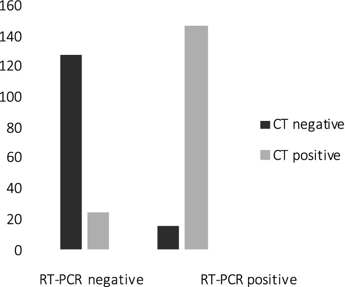
Methods: Three hundred and fourteen patients presented with clinically suspected COVID-19, from March 3 to 23, 2020, were evaluated with PaO2/FIO2 ratio from arterial blood gas, RT-PCR assay from nasopharyngeal swab sample and chest CT. Patients were classified as COVID-19 negative and COVID-19 positive according to RT-PCR results, considered as a reference. Images were independently evaluated by two radiologists blinded to the RT-PCR results and classified as "CT positive" or "CT negative" for COVID-19, according to CT findings.
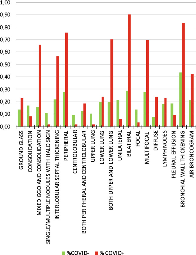
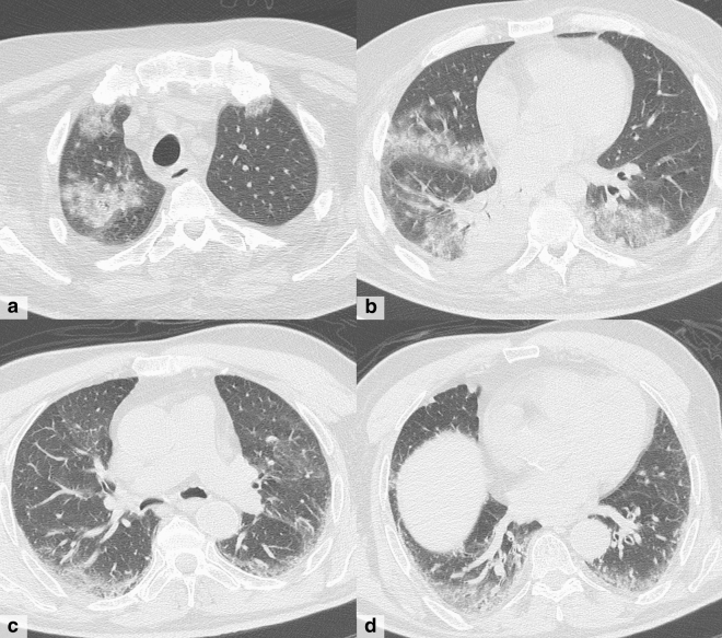
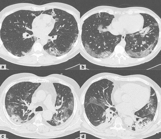
Results: According to RT-PCR results, 152 patients were COVID-19 negative (48%) and 162 were COVID-19 positive (52%). We found substantial agreement between RT-PCR results and CT findings (p < 0.000001), as well as an almost perfect agreement between the two readers. Mixed GGO and consolidation pattern with peripheral and bilateral distribution, multifocal or diffuse abnormalities localized in both upper lung and lower lung, in association with interlobular septal thickening, bronchial wall thickening and air bronchogram, showed higher frequency in COVID-positive patients. We also found a significant correlation between CT findings and patient's oxygenation status expressed by PaO2/FIO2 ratio.
Conclusion: Chest CT has a useful role in the early detection and in patient management of COVID-19 pneumonia in a pandemic. It helps in identifying suspected patients, cutting off the route of transmission and avoiding further spread of infection.
Keywords: COVID-19; Chest CT; Emergency radiology; Pandemic; Pneumonia.
Conflict of interest statement
The authors declare that they have no conflict of interest.
Figures
References
-
- Gazzetta Ufficiale. https://www.gazzettaufficiale.it/eli/id/2014/5/31/14G00093/sg. Accessed 26 July 2019
-
- Evaluating the accuracy of different respiratory specimens in the laboratory diagnosis and monitoring the viral shedding of 2019-nCoV infections | medRxiv. https://www.medrxiv.org/content/10.1101/2020.02.11.20021493v2. Accessed 7 Apr 2020 - DOI
-
- WHO-2019-nCoV-SurveillanceGuidance-2020.6-eng.pdf
MeSH terms
LinkOut - more resources
Full Text Sources
Medical
