

Implementation of a Deep Learning-Based Computer-Aided Detection System for the Interpretation of Chest Radiographs in Patients Suspected for COVID-19
- PMID: 32729263
- PMCID: PMC7458860
- DOI: 10.3348/kjr.2020.0536
Implementation of a Deep Learning-Based Computer-Aided Detection System for the Interpretation of Chest Radiographs in Patients Suspected for COVID-19
Abstract
Objective: To describe the experience of implementing a deep learning-based computer-aided detection (CAD) system for the interpretation of chest X-ray radiographs (CXR) of suspected coronavirus disease (COVID-19) patients and investigate the diagnostic performance of CXR interpretation with CAD assistance.
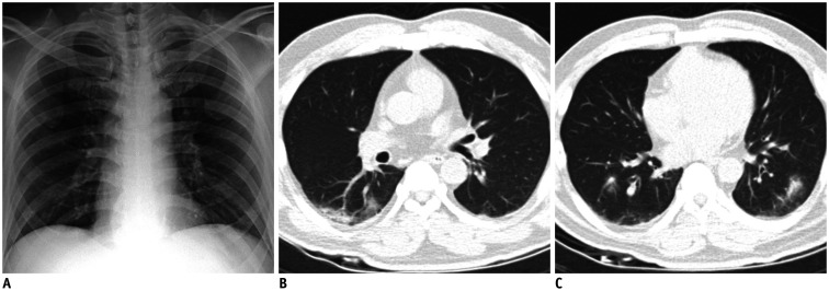
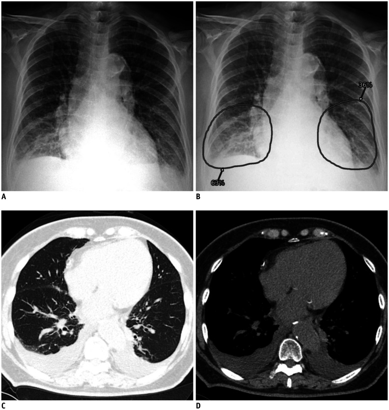
Materials and methods: In this single-center retrospective study, initial CXR of patients with suspected or confirmed COVID-19 were investigated. A commercialized deep learning-based CAD system that can identify various abnormalities on CXR was implemented for the interpretation of CXR in daily practice. The diagnostic performance of radiologists with CAD assistance were evaluated based on two different reference standards: 1) real-time reverse transcriptase-polymerase chain reaction (rRT-PCR) results for COVID-19 and 2) pulmonary abnormality suggesting pneumonia on chest CT. The turnaround times (TATs) of radiology reports for CXR and rRT-PCR results were also evaluated.
Results: Among 332 patients (male:female, 173:159; mean age, 57 years) with available rRT-PCR results, 16 patients (4.8%) were diagnosed with COVID-19. Using CXR, radiologists with CAD assistance identified rRT-PCR positive COVID-19 patients with sensitivity and specificity of 68.8% and 66.7%, respectively. Among 119 patients (male:female, 75:44; mean age, 69 years) with available chest CTs, radiologists assisted by CAD reported pneumonia on CXR with a sensitivity of 81.5% and a specificity of 72.3%. The TATs of CXR reports were significantly shorter than those of rRT-PCR results (median 51 vs. 507 minutes; p < 0.001).
Conclusion: Radiologists with CAD assistance could identify patients with rRT-PCR-positive COVID-19 or pneumonia on CXR with a reasonably acceptable performance. In patients suspected with COVID-19, CXR had much faster TATs than rRT-PCRs.
Keywords: COVID-19; COVID-19 diagnostic testing; Deep learning; Pneumonia; Radiography, thoracic.
Copyright © 2020 The Korean Society of Radiology.
Conflict of interest statement
Eui Jin Hwang, Hyungjin Kim, and Chang Min Park report research grants from the Lunit Inc., outside the present study. Jin Mo Goo report research grant from the INFINITT Healthcare, outside the present study.
Figures

References
-
- National Authorities. Coronavirus disease (COVID-19). Situation report-127. World Health Organization; 2020. [Accessed May 26, 2020]. Available at: https://www.who.int/docs/default-source/coronaviruse/situation-reports/2....
-
- Interim guidelines for collecting, handling, and testing clinical specimens from persons for coronavirus disease 2019 (COVID-19) Centers for Disease Control and Prevention Web site; [Accessed May 26, 2020]. https://www.cdc.gov/coronavirus/2019-nCoV/lab/guidelines-clinical-specim.... Published May 22, 2020.
Publication types
MeSH terms
Grants and funding
LinkOut - more resources
Full Text Sources
Medical
Research Materials
Miscellaneous
