. 2020 Jul 30:9:e58625.
doi: 10.7554/eLife.58625.

Keratinocytes contribute to normal cold and heat sensation

Affiliations

Keratinocytes contribute to normal cold and heat sensation

Katelyn E Sadler et al. Elife. .

Abstract

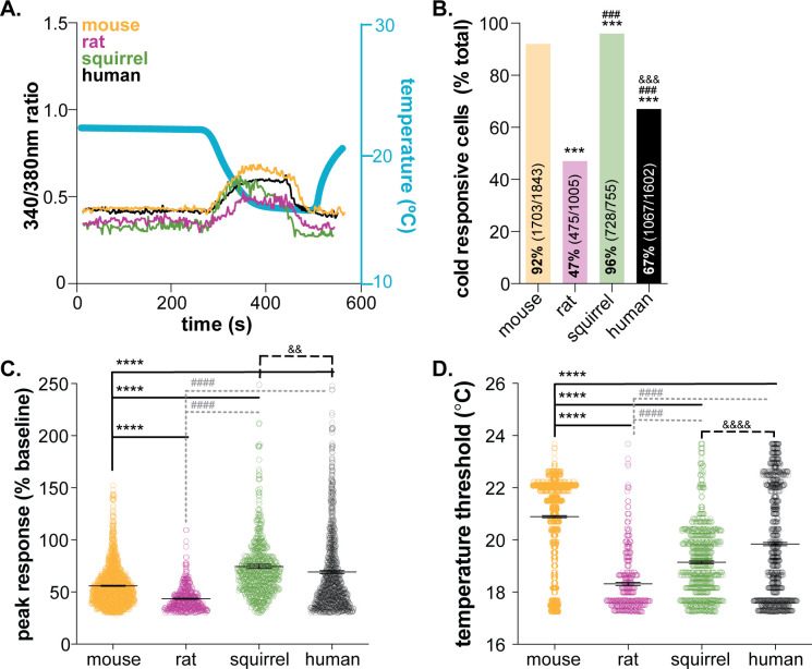
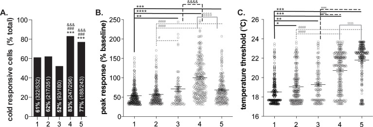
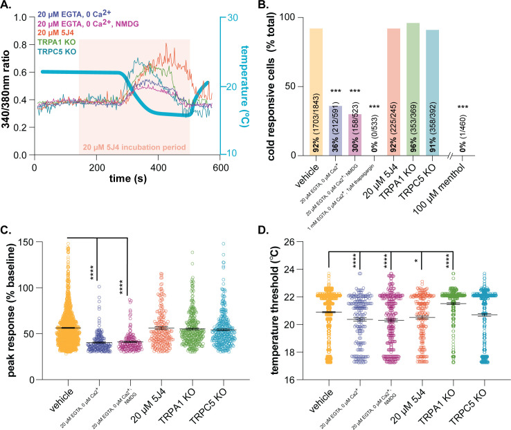
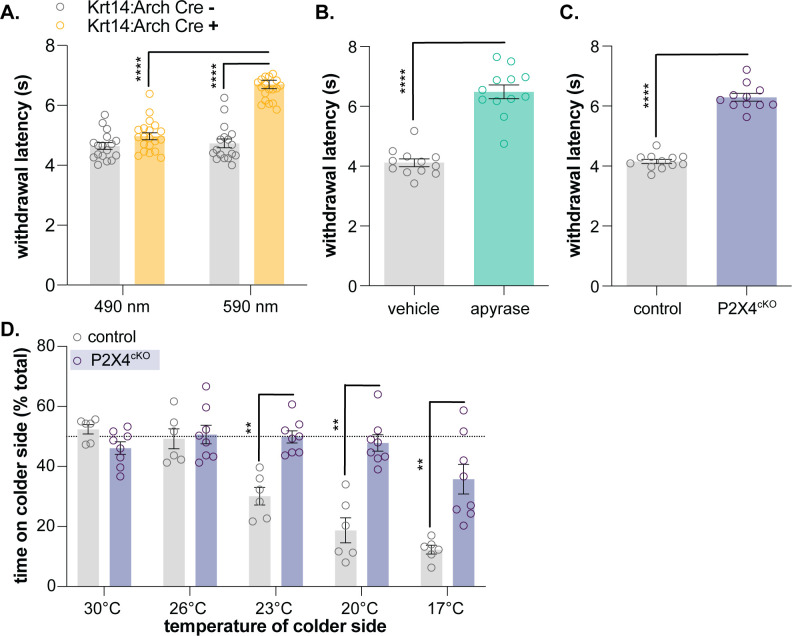
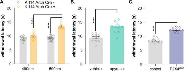
Keratinocytes are the most abundant cell type in the epidermis, the most superficial layer of skin. Historically, epidermal-innervating sensory neurons were thought to be the exclusive detectors and transmitters of environmental stimuli. However, recent work from our lab (Moehring et al., 2018) and others (Baumbauer et al., 2015) has demonstrated that keratinocytes are also critical for normal mechanotransduction and mechanically-evoked behavioral responses in mice. Here, we asked whether keratinocyte activity is also required for normal cold and heat sensation. Using calcium imaging, we determined that keratinocyte cold activity is conserved across mammalian species and requires the release of intracellular calcium through one or more unknown cold-sensitive proteins. Both epidermal cell optogenetic inhibition and interruption of ATP-P2X4 signaling reduced reflexive behavioral responses to cold and heat stimuli. Based on these data and our previous findings, keratinocyte purinergic signaling is a modality-conserved amplification system that is required for normal somatosensation in vivo.

Keywords: ATP; P2X4; cold; heat; human; keratinocyte; mouse; neuroscience; rat.


Conflict of interest statement

KS, FM, CS No competing interests declared

Figures

Figure 1.
Figure 1.. Mammalian keratinocytes respond to temperature decreases.
In vitro calcium imaging was performed on primary cultured keratinocytes from the glabrous hindpaw skin of C57BL/6 mice, Sprague Dawley rats, and 13-lined ground squirrels and human breast skin. (A) Representative keratinocytes from all species exhibited a calcium transient upon extracellular buffer cooling (~23°C to 12°C). (B) The greatest proportion of cold-responsive cells was observed in hibernating 13-lined ground squirrel samples. The vast majority of mouse keratinocytes also responded to decreasing temperatures; fewer rat and human keratinocytes responded to cold (Chi-square p<0.0001; Fisher’s Exact tests: ***p<0.001 vs. mouse; ### p<0.001 vs. rat; &&& p<0.001 vs. squirrel; n = 28 mice, four rats, four squirrels, five humans). (C) The peak calcium response to decreasing temperatures varied between species; hibernating 13-lined ground squirrel keratinocytes exhibited the largest calcium transients (75% increase over baseline) and Sprague Dawley rats exhibited the smallest (44% increase over baseline; mouse: 56% increase; human: 69% increase; 1-way ANOVA p<0.0001; Bonferroni’s multiple comparisons: ****p<0.0001 vs. mouse; #### p<0.0001 vs. rat, && p=0.0084 vs. squirrel). (D) The temperature at which keratinocytes responded to cold (>30% increase in Δ340/380) differed between species. Mean temperature thresholds (°C) for mouse: 20.9, rat: 18.3, squirrel: 19.1, human: 19.8 (Kruskal-Wallis test p<0.0001; Dunn’s multiple comparisons: ****p<0.0001 vs. mouse, #### p<0.0001 vs. rat, &&&& p<0.0001 vs. squirrel).
Figure 1—figure supplement 1.
Figure 1—figure supplement 1.. Variability in human keratinocyte responses to temperature decreases.
In vitro calcium imaging was performed on primary human breast skin keratinocytes isolated from females of the following ages (years): 1: 41, 2: 50, 3: 54, 4: 52, 5: 49. (A).). Samples 4 and 5 contained the most cold-responsive cells (Chi-square: p<0.0001; Fisher’s Exact tests: ***p<0.001 vs. sample 1, ### p<0.001 vs. sample 2, &&& p<0.001 vs. sample 3). (B).) Peak calcium responses differed between the samples; keratinocytes from sample four exhibited the largest calcium response during cold stimulation (100% increase over baseline. Mean increase for sample 1: 54%, 2: 57%, 3: 71%, 5: 68% (1-way ANOVA p<0.0001; Bonferroni’s multiple comparisons: **p<0.01, ***p<0.001, ****p<0.0001 vs. 1; # p<0.05, #### p<0.0001 vs. 2; &&&& p<0.0001 vs. 3; $$$$ p<0.0001 vs. 4). (C).) The temperature at which keratinocytes responded to cold (>30% increase in Δ340/380) differed between the samples. Mean temperature thresholds (°C) for sample 1: 18.5, 2: 19.1, 3: 19.2, 4: 20.7, 5: 21.8 (Kruskal-Wallis test p<0.0001; Dunn’s multiple comparisons: *p<0.05, **p<0.01, ***p<0.001 vs. 1; ### p<0.001 vs. 2; &&&& p<0.0001 vs. 3; $$$$ p<0.0001 vs. 4).
Figure 2.
Figure 2.. Characterizing murine cold sensor(s) in keratinocytes.
(A).) In order to characterize the proteins involved in cold transduction, cold-induced calcium transients were measured in keratinocytes exposed to extracellular buffer containing different ionic concentrations or pharmacological agents, or in keratinocytes from transgenic mice. (B).) Extracellular calcium chelation (EGTA, 0 µM Ca2+) decreases the proportion of keratinocytes that respond to cold; substituting NMDG for extracellular sodium did not further decrease the percentage of cold-responsive cells. Cold responses were only abolished when endoplasmic reticulum Ca2+ stores were depleted and unable to be refilled with extracellular sources (EGTA, 0 µM Ca2+, thapsigargin). CRAC channel inhibition (5J4) did not decrease the percentage of cold-responsive cells, and similar proportions of keratinocytes from C57BL/6, global TRPA1, and global TRPC5 knockout mice responded to decreasing buffer temperature (Chi square p<0.0001; Fisher’s Exact tests ***p<0.001 vs. vehicle; n ≥ 4). (C).) Peak calcium responses to cold were lower in the absence of extracellular calcium; responses were unaltered in other conditions (1-way ANOVA p<0.0001; Bonferroni’s multiple comparisons: ****p<0.0001 vs. vehicle, #### p<0.0001 vs. EGTA, 0 µM Ca2+, &&&& p<0.0001 vs. EGTA, 0 µM Ca2+, NMDG). (D).) Altering extracellular buffer contents increased or decreased the temperature at which keratinocytes responded to cold by ≤0.6°C. Mean temperature thresholds (°C) for vehicle: 20.9, EGTA, 0 µM Ca2+,±NMDG: 20.3, 20 µM 5J4: 20.5, TRPA1 KO: 21.5, TRPC5 KO: 20.7 (Kruskal-Wallis test p<0.0001; Dunn’s multiple comparisons: *p<0.05, ****p<0.0001 vs. vehicle).
Figure 3.
Figure 3.. Keratinocyte-to-sensory neuron signaling is required for normal cold sensation.
(A) 590 nm light exposure increases the withdrawal latency of Krt14:Arch Cre+ mice during plantar dry ice stimulation (2-way ANOVA main effects of light, genotype, and light x genotype interaction p<0.0001; Bonferroni’s multiple comparisons: ****p<0.0001 Krt14:Arch Cre+ 490 vs. 590 nm, 590 nm Krt14:Arch Cre+ vs. Cre-; n = 17–20) (B) Intraplantar administration of apyrase (0.4 units; catalyzes ATP hydrolysis) increases the withdrawal latency of C57BL/6 mice during plantar dry ice stimulation (unpaired t-test ****p<0.0001; n = 12). (C) Animals lacking sensory neuron P2X4 receptors (P2X4cKO) exhibit increased withdrawal latencies to plantar dry ice stimulation (unpaired t-test ****p<0.0001; n = 11–12). (D) In a two-temperature preference test, P2X4cKO mice spend more time on innocuous cold surfaces than their P2X4 expressing littermates (2-way ANOVA main effects of temperature, genotype, and temperature x genotype interaction p<0.0001; Bonferroni’s multiple comparisons: **p<0.01 P2X4cKO vs. control; n = 6–8).
Figure 4.
Figure 4.. Keratinocyte-to-sensory neuron signaling is required for normal heat sensation.
(A) 590 nm light exposure increases the withdrawal latency of Krt14:Arch Cre+ mice during plantar radiant heat stimulation (2-way ANOVA main effects of light, genotype, and light x genotype interaction p<0.0001; Bonferroni’s multiple comparisons: ****p<0.0001 Krt14:Arch Cre+ 490 vs. 590 nm, 590 nm Krt14:Arch Cre+ vs. Cre-; n = 17–20). (B) Intraplantar administration of apyrase (0.4 units; catalyzes ATP hydrolysis) increases the withdrawal latency of C57BL/6 mice during plantar radiant stimulation (unpaired t-test ****p<0.0001; n = 10). (C) Animals lacking sensory neuron P2X4 receptors (P2X4cKO) exhibit increased withdrawal latencies to plantar radiant heat stimulation (unpaired t-test ****p<0.0001; n = 11–12).

References

    1. Askwith CC, Benson CJ, Welsh MJ, Snyder PM. DEG/ENaC ion channels involved in sensory transduction are modulated by cold temperature. PNAS. 2001;98:6459–6463. doi: 10.1073/pnas.111155398. - DOI - PMC - PubMed
    1. Baumbauer KM, DeBerry JJ, Adelman PC, Miller RH, Hachisuka J, Lee KH, Ross SE, Koerber HR, Davis BM, Albers KM. Keratinocytes can modulate and directly initiate nociceptive responses. eLife. 2015;4:e09674. doi: 10.7554/eLife.09674. - DOI - PMC - PubMed
    1. Bautista DM, Jordt SE, Nikai T, Tsuruda PR, Read AJ, Poblete J, Yamoah EN, Basbaum AI, Julius D. TRPA1 mediates the inflammatory actions of environmental irritants and proalgesic agents. Cell. 2006;124:1269–1282. doi: 10.1016/j.cell.2006.02.023. - DOI - PubMed
    1. Beck B, Zholos A, Sydorenko V, Roudbaraki M, Lehen'kyi V, Bordat P, Prevarskaya N, Skryma R. TRPC7 is a receptor-operated DAG-activated channel in human keratinocytes. Journal of Investigative Dermatology. 2006;126:1982–1993. doi: 10.1038/sj.jid.5700352. - DOI - PubMed
    1. Bidaux G, Borowiec AS, Gordienko D, Beck B, Shapovalov GG, Lemonnier L, Flourakis M, Vandenberghe M, Slomianny C, Dewailly E, Delcourt P, Desruelles E, Ritaine A, Polakowska R, Lesage J, Chami M, Skryma R, Prevarskaya N. Epidermal TRPM8 channel isoform controls the balance between keratinocyte proliferation and differentiation in a cold-dependent manner. PNAS. 2015;112:E3345–E3354. doi: 10.1073/pnas.1423357112. - DOI - PMC - PubMed

Publication types

MeSH terms