Review
. 2021 Apr 1;101(2):427-493.
doi: 10.1152/physrev.00043.2019. Epub 2020 Jul 30.

Role of O-Linked N-Acetylglucosamine Protein Modification in Cellular (Patho)Physiology

Affiliations
Review

Role of O-Linked N-Acetylglucosamine Protein Modification in Cellular (Patho)Physiology

John C Chatham et al. Physiol Rev. .

Abstract

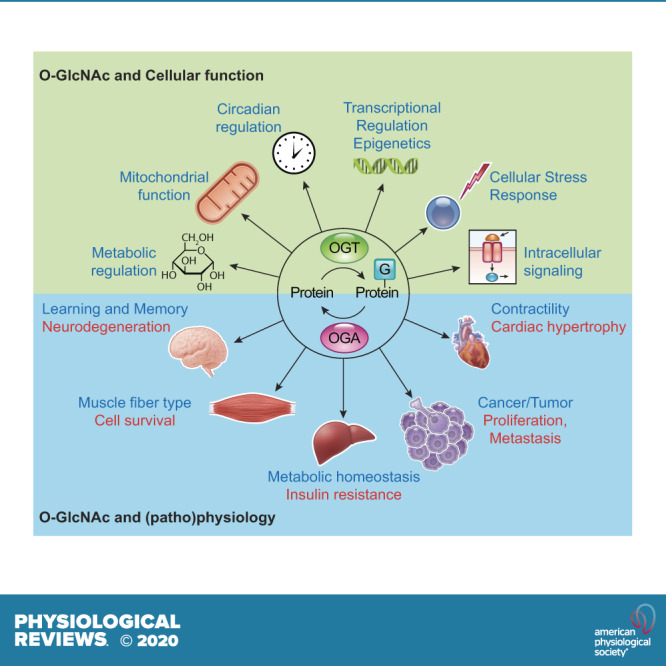
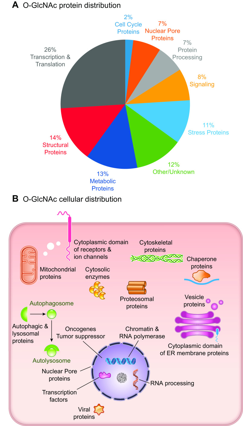
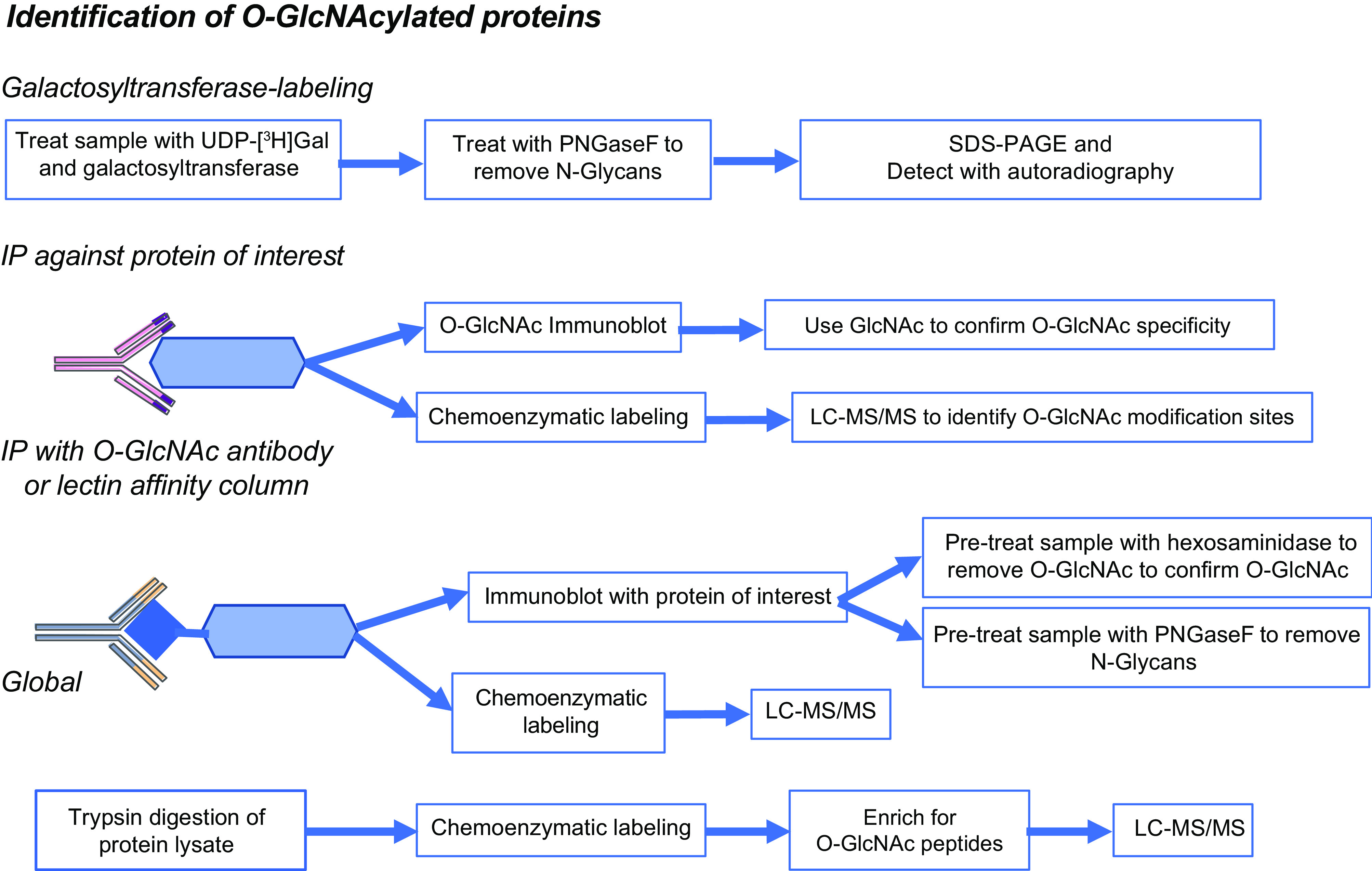
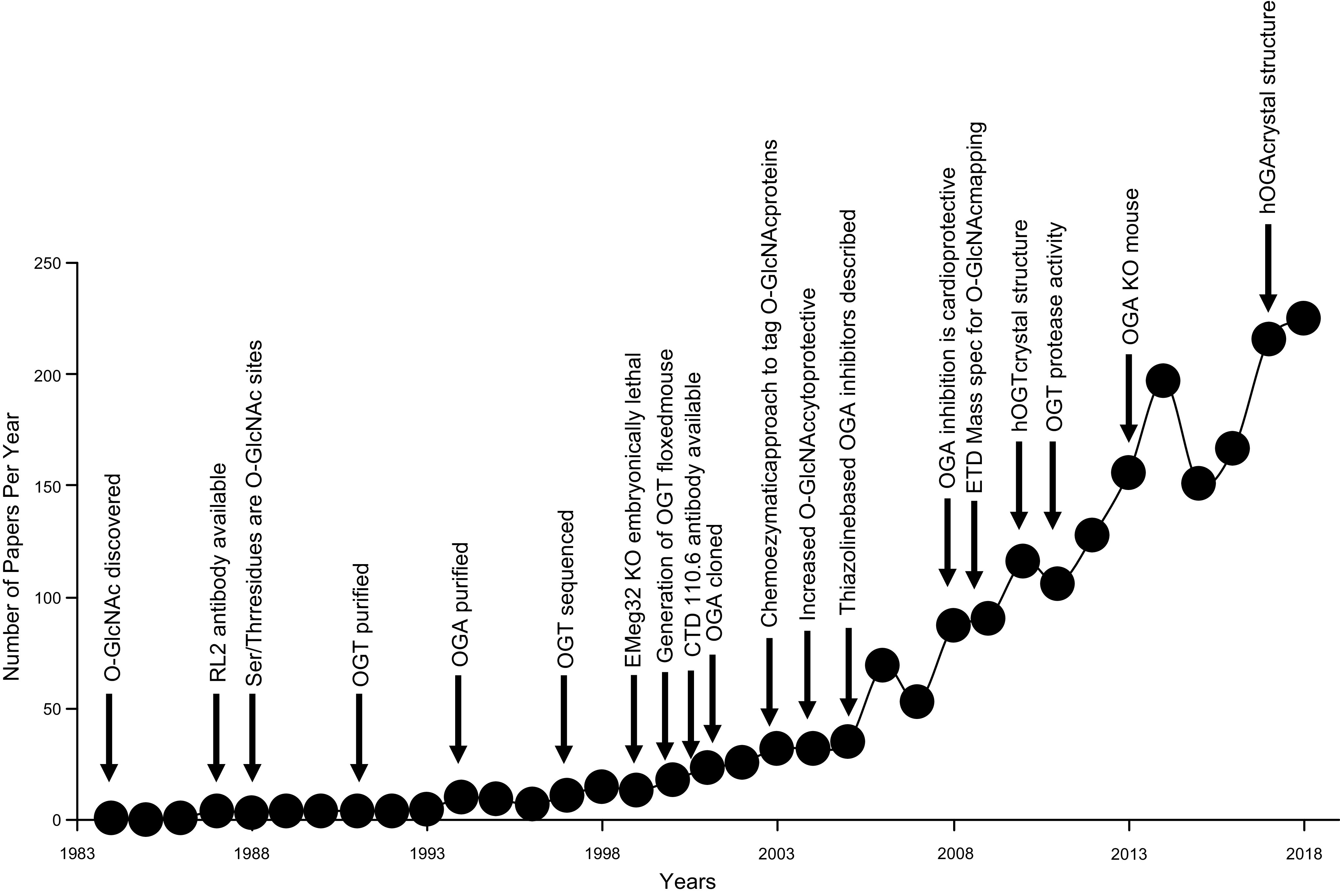
In the mid-1980s, the identification of serine and threonine residues on nuclear and cytoplasmic proteins modified by a N-acetylglucosamine moiety (O-GlcNAc) via an O-linkage overturned the widely held assumption that glycosylation only occurred in the endoplasmic reticulum, Golgi apparatus, and secretory pathways. In contrast to traditional glycosylation, the O-GlcNAc modification does not lead to complex, branched glycan structures and is rapidly cycled on and off proteins by O-GlcNAc transferase (OGT) and O-GlcNAcase (OGA), respectively. Since its discovery, O-GlcNAcylation has been shown to contribute to numerous cellular functions, including signaling, protein localization and stability, transcription, chromatin remodeling, mitochondrial function, and cell survival. Dysregulation in O-GlcNAc cycling has been implicated in the progression of a wide range of diseases, such as diabetes, diabetic complications, cancer, cardiovascular, and neurodegenerative diseases. This review will outline our current understanding of the processes involved in regulating O-GlcNAc turnover, the role of O-GlcNAcylation in regulating cellular physiology, and how dysregulation in O-GlcNAc cycling contributes to pathophysiological processes.

Keywords: calcium; cancer; diabetes; genetics; metabolism.


Figures

None
Graphical abstract
FIGURE 1.
FIGURE 1.
A: O-linked N-acetylglucosamine acylated (O-GlcNAcylated) proteins belong to many different classes of proteins responsible for regulating diverse cellular processes. Some of the largest classes of proteins include those in regulating metabolism, transcription, and translation, as well as structural proteins. B: O-GlcNAcylated proteins are present in numerous cellular compartments, including the nucleus, cytosol, and mitochondria. Cytosolic domains of membrane proteins are also O-GlcNAcylated, as well as proteins involved in autophagy and proteosomal degradation of proteins, chaperone proteins, vesicle proteins, and numerous cytosolic proteins and enzymes. This figure is based, in part, on information presented in Chapter 19, The O-GlcNAc Modification, Essentials in Glycobiology, 3rd ed. (14). ER, endoplasmic reticulum.
FIGURE 2.
FIGURE 2.
Overview of different approaches for the identification of O-linked N-acetylglucosamine acylated (O-GlcNAcylated) proteins, including galactosyltransferase labeling; immunopurification and chemoenzymatic labeling were combined with LC-MS/MS. Further details are found in TABLE 1. IP, immunoprecipitation; UDP, uridine diphosphate-azido-modified galactose.
FIGURE 3.
FIGURE 3.
Number of O-linked N-acetylglucosamine acylated (O-GlcNAc) publications by year annotated by key events in O-GlcNAc biology from its initial discovery. Relevant citations are all included in the main text. CTD, COOH-terminal domain; ETD, electron dissociation transfer hOGA, human O-GlcNAcase; hOGT, human O-GlcNAc transferase; KO, knockout; OGA, O-GlcNAcase; OGT, O-GlcNAc transferase; RL2, rat liver nuclear pore complex.
FIGURE 4.
FIGURE 4.
Schematic of UDP-GlcNAc and O-GlcNAc synthesis. Glucose enters the cell via the glucose transporter system where it is rapidly phosphorylated by hexokinase (HK) and converted to fructose-6-phosphate by phosphoglucoseisomerase (PGI). Fructose-6-phosphate is subsequently metabolized to glucosamine-6-phosphate by l-Glutamine: d-fructose-6-phosphate amidotransferase (GFAT), which requires glutamine. Glucosamine-6-phosphate is converted to N-acetylglucosamine-6-phosphate by glucosamine 6-phosphate N-acetyltransferase (Emeg32), utilizing acetyl-CoA. Phosphoacetylglucosamine mutase (Agm1) converts N-acetylglucosamine-6-phosphate to N-acetylglucosamine-1-phosphate. The synthesis of uridine-diphosphate-N-acetylglucosamine (UDP-GlcNAc) is catalyzed by UDP-N-acetylglucosamine pyrophosphorylase (Uap1), which consumes uridine triphosphate (UTP). UDP-GlcNAc is the substrate for (O-GlcNAc transferase (OGT) leading to the formation of O-linked β-N-acetylglucosamine (O-GlcNAc)-modified proteins. β-N-acetylglucosaminidase (OGA) catalyzes the removal of O-GlcNAc from the proteins. GlcNAc can reenter the HBP via two salvage pathways: 1) via N-acetylglucosamine kinase (NAGK) to generate N-acetylglucosamine 1-phosphate and 2) involving the conversion by N-acetylgalactosamine kinase (GALK2) of N-acetyl-galactosamine to N-acetylgalactosamine 1-phosphate and UDP-N-acetylgalactosamine, with subsequent conversion by an epimerase to UDP-GlcNAc. Glucosamine, which enters the cell via the glucose transport system and can be phosphorylated by hexokinase (HK) to form glucosamine 6-phosphate thereby bypassing GFAT. The kinases that have been identified as regulating GFAT, OGT, and OGA are indicated; additional details may be found in the text (see FIGURE 6 and TABLE 3). AMPK, AMPK-activated protein kinase; CaMKII, calcium/calmodulin (Ca2+/CaM) dependent protein kinase II; CHK1, checkpoint kinase-1; CK2, casein kinase 2; EPI, epimerase; GalNAc, glucosamine fructose-6-phosphate amidotransferase; GFAT, glucosamine fructose-6-phosphate amidotransferase; GlcNAc, N-acetylglucosamine; GlcNAc-1P, N-acetylglucosamine-1-phosphate; GSK3β, glycogen synthase kinase 3b; HEX, hexokinase; IRS1, insulin receptor substrate-1; PPi, pyrophosphate; UDP-GalNAc, uridine diphosphate N-acetylgalactosamine.
FIGURE 5.
FIGURE 5.
Regulation of glucosamine fructose-6-phosphate amidotransferase (GFAT). GFAT activity is regulated at several levels including substrate availability, feedback inhibition by uridine diphosphate N-acetylglucosamine (UDP-GlcNAc) and glucosamine-6-phosphate (GlcN-6-P), as well as post-translational modifications by phosphorylation, acetylation, succinylation, and ubiquitination. The only kinases that have been identified as phosphorylating GFAT are PKA, AMPK, and calcium/calmodulin (Ca2+/CaM) dependent protein kinase II (CaMKII). Sp1, ATF4, and XBP1s have been shown to regulate GFAT at the transcriptional level. The database phosphosite.org and the citations contained therein were used to identify known posttranslational modification sites of GFAT (196). ATF4, activating transcription factor 4; Lys, lysine; Sp1, specificity protein 1; XBP1s, X-box binding protein 1.
FIGURE 6.
FIGURE 6.
Structure and posttranslational modifications of human OGT (A) and OGA (B). Phosphorylation sites with identified kinases are shown as red circles, and all known phosphorylation sites are listed below the figures. O-GlcNAc sites are shown as blue squares, and acetylation sites are shown as green circles. AT, acetyl transferase; CaMKII, calcium/calmodulin (Ca2+/CaM) dependent protein kinase II; CD, catalytic domain; CHK1, checkpoint kinase-1; GSK3β, glycogen synthase kinase 3β; hOGT, human O-GlcNAc transferase; hOGA, huma O-GlcNAcase; Int-D, intervening domain; IRS1, insulin receptor substrate-1; LC, low complexity region; NLS, nuclear localization sequence; TPR, tetratricopeptide repeats. The databases https://www.phosphosite.org and http://www.phosphonet.ca/, and the citations contained therein were used to for known phosphorylation sites and acetylation modification sites(196). Citations for sites where kinases have been identified are included in the text; additional resources include Lundby, et al. (235), Levine and Walker (232) and Roth et al. (236). Other PTMs not shown include ubiquitination and sumoylation.
FIGURE 7.
FIGURE 7.
Extensive crosstalk between phosphorylation and O-GlcNAcylation. A, i: example of phosphorylation and O-GlcNAcylation of the same or adjacent sites that prevent simultaneous modifications. and A, ii: modification by both phosphorylation and O-GlcNAcylation, as well as O-GlcNAcylation enhancing phosphorylation via for example increased binding to adaptor proteins. B, i: example of a kinase that is activated when phosphorylated and inactivated by O-GlcNAcylation. B, ii: a kinase that can be modified by both O-GlcNAc and phosphorylation involving complex interactions between OGT/OGA and kinases/phosphatases. C: O-GlcNAcylation also regulates phosphorylation via modification of phosphatases, and OGT activity is directly regulated by kinase phosphorylation. GSK3β, glycogen synthase kinase 3β; OGA, O-GlcNAcase; OGT, O-GlcNAc transferase; PPT, protein phosphatase.
FIGURE 8.
FIGURE 8.
O-linked β-N-acetylglucosamine (O-GlcNAc) regulation of gene expression and epigenetics. A: examples of transcriptional regulation mediated by O-GlcNAc with additional details and references highlighted in the text. These examples include both inhibitory and activating roles of O-GlcNAc (blue square with G). A, I: for the transcription factor Sp1, one O-GlcNAc modification blocks an activation site to inhibit transcription, while a different O-GlcNAc site inhibits proteasomal degradation increasing transcription. A, II: a separate set of examples of this dual role of O-GlcNAc-mediated regulation is at ChREBP, which, under normal glucose levels, leads to O-GlcNAc modification (blue square) to enhance 14-3-3 binding decreasing transcriptional activity, while under high glucose levels, additional posttranslational regulation by phosphorylation (red circle with P) leads to a different O-GlcNAc modification and augments activity. A, III: multiple roles are shown for both direct O-GlcNAc modification of RNA polymerase II (RNAP II), as well as the contribution of uridine diphosphate-azido-modified galactose (UDP)-GlcNAc hydrolysis to enhance transcriptional activity. A, IV: multiple roles are shown for different O-GlcNAc modifications to either inhibit ubiquitination (gray square with U) to increase complex stability or other modifications that may impact nuclear cytoplasmic import/export of cargo (e.g., RNA). B: examples of epigenetic regulation mediated by O-GlcNAc with additional details and references highlighted in the text. B, I: highlights the multiple direct (i.e., O-GlcNAc of histone proteins) and indirect (i.e. interaction and modification of other histone modifiers), such as EZH2 to impact histone methylation (black square with M) and SIN3a to impact histone acetylation (purple square with A). B, II: highlights the interaction of O-GlcNAc transferase (OGT) with the ten-eleven translocation (TET) enzymes impacting DNA 5 hydroxymethylation (5hmC) which, in turn, can lead to DNA demethylation. B, III: a few examples are highlighted from the text of noncoding RNA [ncRNA; e.g., long-noncoding RNA (lncRNA) and microRNA (miR)] regulation of O-GlcNAc enzymes.
FIGURE 9.
FIGURE 9.
Examples of O-linked β-N-acetylglucosamine (O-GlcNAc) regulation of cellular signaling pathways. A: Insulin signaling. On initiation, in response to insulin, there is autophosphorylation of the insulin receptor (IR) and subsequent phosphorylation and activation of Akt, FOXO and GSK3β and their downstream signaling pathways. On termination, insulin triggers rapid translocation of O-GlcNAc transferase (OGT) from the nucleus to the plasma membrane, where it is phosphorylated by the IR, which increases OGT activity, leading to the subsequent O-GlcNAcylation of insulin receptor substrate 1 (IRS1), AKT, FOXO, and GSK3β, thereby attenuating activity at multiple steps in the insulin signaling cascade. The protein tyrosine phosphatase 1B (PTP1B) is responsible for decreasing phosphorylation of IR and attenuating insulin signaling. It is also O-GlcNAcylated, which increases its activity contributing to a further reduction in insulin signaling. It has been reported that translocation of OGT to the plasma membrane is via binding to PIP3 (242), which is generated in response to activation of insulin signaling; however, to date, structural studies have not revealed a phosphatidylinositol-3-phosphate (PIP3)-binding motif in OGT(90). B: Calcium signaling. Intracellular Ca2+ increases in response to diverse number of agonists, either as a result of Ca2+ release from ER/SR alone or via activation of plasma membrane Ca2+ channels as a result of ER/SR Ca2+ release (store-operated Ca2+ entry, SOCE). This increase in Ca2+ leads to activation of calmodulin (CaM), resulting in the phosphorylation and activation of both CaMKII and IV, which are responsible for regulation of numerous cellular processes. CaMKII also phosphorylates OGT, increasing its activity, and in a feedback manner, OGT O-GlcNAcylates CaMKII/IV reduces their activities. Ca2+-CaM also activates the calcium-dependent phosphatase calcineurin, which is responsible for regulating diverse cellular processes, and it has been reported that increased OGT expression and O-GlcNAcylation is sufficient to activate calcineurin-mediated transcription pathways (298). Influx of extracellular Ca2+ has been shown to contribute to the stress-induced increases in O-GlcNAc levels (312). Therefore, it is possible, that Ca2+-CaM and/or calcineurin regulate OGT (or OGA) activities; however, this has yet to be demonstrated experimentally. Multiple contractile proteins are O-GlcNAc modified (TABLE 2), and the increases in O-GlcNAc levels that can occur in diseases, such as diabetes, reduces Ca2+ sensitivity of some contractile proteins (147, 150). Phospholamban (PLB) and SERCA are responsible, in part, for the reuptake of Ca2+ into the sarcoplasmic reticulum (SR), contributing to muscle relaxation. Increases in O-GlcNAc levels decrease the activities of both PLB and SERCA directly or indirectly, which could be a contributing factor to impaired myocardial relaxation (i.e., diastolic function) that occurs with diabetes. The endoplasmic reticulum (ER)/SR transmembrane protein STIM1, which plays a key role in regulating SOCE, is also a target for O-GlcNAcylation and increases in O-GlcNAc levels impairs its function and attenuates SOCE (401). Increasing O-GlcNAc levels attenuates mitochondrial Ca2+ overload, although the precise mechanisms are unclear(163, 402). CaMKII phosphorylation of the mitochondrial Ca2+ uniporter (MCU) potentiates mitochondrial Ca2+ overload (403); therefore, as O-GlcNAcylation of CaMKII decreases its activity, this may represent a potential protective mechanism. However, others have questioned the role of CaMKII in the regulation of mitochondrial Ca2+ uptake by the MCU (404).
FIGURE 10.
FIGURE 10.
Metabolism and mitochondrial O-GlcNAcylation. A: majority of enzymes in glycolysis and glucose metabolism are targets for O-GlcNAcylation (418). ALD, aldolase; ENO, enolase; GAPDH, glyceraldehyde-6-phosphate dehydrogenase; GFAT, glutamine fructose-6phosphate amidotransferase; GP, glycogen phosphorylase; G6PDH, glucose-6-phosphate dehydrogenase; GS, glycogen synthase; HEX, hexokinase; LDH, lactate dehydrogenase; PFK, phosphofructokinase; PDC, pyruvate dehydrogenase complex; PGI, phosphoglucoisomerase; PGK, phosphoglycerate kinase; PGM phosphoglucomutase; PK, pyruvate kinase; UDP-GP, UDP-glucose pyrophosphorylase. B: wide range of different types of mitochondrial proteins that are O-GlcNAcylated. C: map of the O-GlcNAc modification sites on proteins that are central to the regulation of energy metabolism and mitochondrial function. Data largely based on a number of recent proteomic studies (138,139). Blue squares denote O-GlcNAc, whereas numbers inside blue squares indicate number of O-GlcNAc sites on the protein.
FIGURE 11.
FIGURE 11.
O-linked β-N-acetylglucosamine (O-GlcNAc) regulation of cell survival and autophagy. A role of O-GlcNAc in cell survival is evidenced by O-GlcNAc transferase (OGT) and O-GlcNAcase (OGA) knockout/knockdown and overexpression studies, although the identities of specific modified proteins in promoting cell death or survival are largely unclear. Among others, AKT, NF-κB, and c-Fos have been shown to be O-GlcNAcylated, and their O-GlcNAcylation is associated with cell death processes. In terms of pathology, Alzheimer’s disease-associated tau phosphorylation has been shown to be attenuated by enhancement of its O-GlcNAcylation. Although there are also studies demonstrating that increased O-GlcNAcylation can enhance proteotoxicity, conceivably by altering the degradation of O-GlcNAcylated forms of specific proteins. The involvement of O-GlcNAcylation in autophagy is best demonstrated in studies on SNAP29 and GRASP55, whose O-GlcNAcylation attenuates autophagic flux.

References

    1. Varki A, Kornfeld S. Historical background and overview. In: Essentials of Glycobiology (3rd ed.), edited by Varki A, Cummings RD, Esko JD, Stanley P, Hart GW, Aebi M, Darvill AG, Kinoshita T, Packer NH, Prestegard JH, Schnaar RL, Seeberger PH.. Cold Spring Harbor, NY: Cold Spring Harbor Laboratory Press, NY, 2015. - PubMed
    1. Torres CR, Hart GW. Topography and polypeptide distribution of terminal N-acetylglucosamine residues on the surfaces of intact lymphocytes. Evidence for O-linked GlcNAc. J Biol Chem 259: 3308–3317, 1984. - PubMed
    1. Holt GD, Hart GW. The subcellular distribution of terminal N-acetylglucosamine moieties. Localization of a novel protein-saccharide linkage, O-linked GlcNAc. J Bioll Chem 261: 8049–8057, 1986. - PubMed
    1. Snow CM, Senior A, Gerace L. Monoclonal antibodies identify a group of nuclear pore complex glycoproteins. J Cell Biol 104: 1143–1156, 1987. doi:10.1083/jcb.104.5.1143. - DOI - PMC - PubMed
    1. Hanover JA, Cohen CK, Willingham MC, Park MK. O-linked N-acetylglucosamine is attached to proteins of the nuclear pore. Evidence for cytoplasmic and nucleoplasmic glycoproteins. J Biol Chem 262: 9887–9894, 1987. - PubMed

Publication types

MeSH terms

Substances

LinkOut - more resources