. 2020 Jul 30;15(7):e0236185.
doi: 10.1371/journal.pone.0236185. eCollection 2020.

mCherry fusions enable the subcellular localization of periplasmic and cytoplasmic proteins in Xanthomonas sp

Affiliations

mCherry fusions enable the subcellular localization of periplasmic and cytoplasmic proteins in Xanthomonas sp

Michelle Mendonça Pena et al. PLoS One. .

Abstract

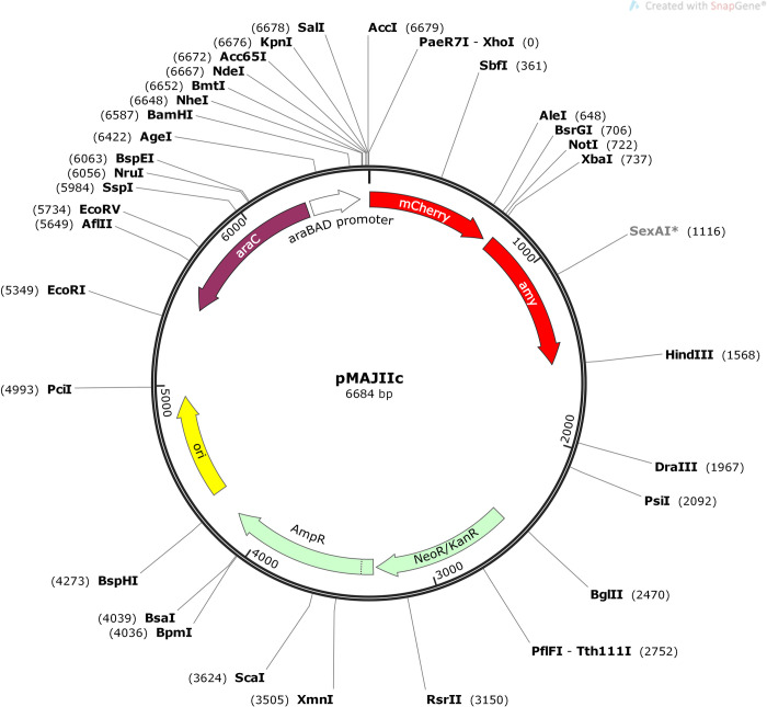
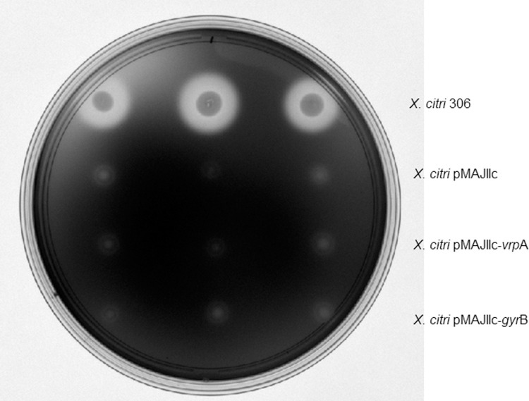
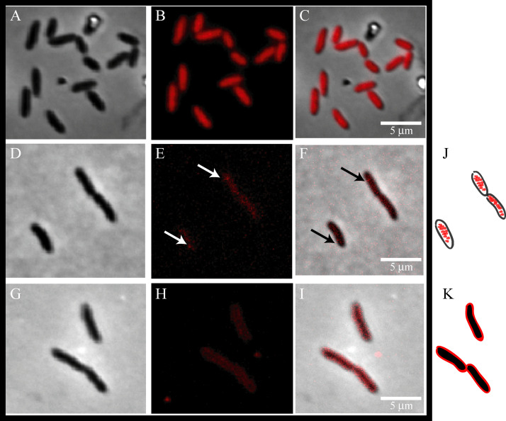
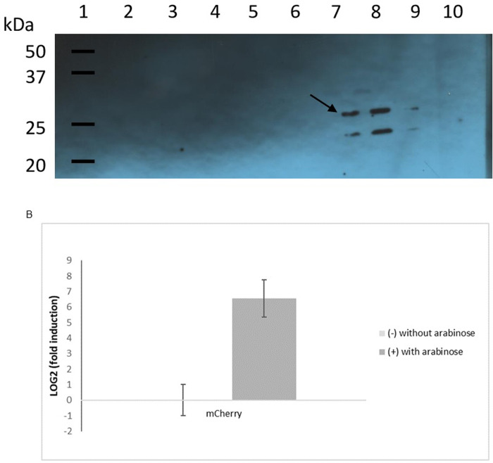
Fluorescent markers are a powerful tool and have been widely applied in biology for different purposes. The genome sequence of Xanthomonas citri subsp. citri (X. citri) revealed that approximately 30% of the genes encoded hypothetical proteins, some of which could play an important role in the success of plant-pathogen interaction and disease triggering. Therefore, revealing their functions is an important strategy to understand the bacterium pathways and mechanisms involved in plant-host interaction. The elucidation of protein function is not a trivial task, but the identification of the subcellular localization of a protein is key to understanding its function. We have constructed an integrative vector, pMAJIIc, under the control of the arabinose promoter, which allows the inducible expression of red fluorescent protein (mCherry) fusions in X. citri, suitable for subcellular localization of target proteins. Fluorescence microscopy was used to track the localization of VrpA protein, which was visualized surrounding the bacterial outer membrane, and the GyrB protein, which showed a diffused cytoplasmic localization, sometimes with dots accumulated near the cellular poles. The integration of the vector into the amy locus of X. citri did not affect bacterial virulence. The vector could be stably maintained in X. citri, and the disruption of the α-amylase gene provided an ease screening method for the selection of the transformant colonies. The results demonstrate that the mCherry-containing vector here described is a powerful tool for bacterial protein localization in cytoplasmic and periplasmic environments.


Conflict of interest statement

The authors have declared that no competing interests exist.

Figures

Fig 1
Fig 1. Map of the integrative vector pMAJIIc.
Ori, replication origin; ap, neo and kan, ampicillin, neomycin and kanamycin resistance, respectively; amy, amy DNA fragment (106–912) of X. citri; mCherry, fluorescent protein; araC, the arabinose repressor; pBAD the arabinose promoter. The polylinker region for in-frame ligation of ORFs/genes to the 5’-end of mCherry, which allows the expression of protein fusions having C-terminal mCherry, is composed of unique sites for NheI, NdeI, KpnI, SalI and XhoI. NotI and XbaI sites, can be used for cloning intended for the expression of proteins carrying N-terminal mCherry fusions. Unique restriction sites are shown in bold letters. The nucleotide sequence of pMAJIIc plasmid was deposited in the GenBank database under the accession number MT119765.
Fig 2
Fig 2. Starch degradation analyses.
Halos corresponding to starch degradation were visualized by exposing the medium to iodine vapor. X. citri 306 produces a clear transparent halo (top), while mutants in which plasmids have integrated into the amy locus are unable to degrade the polymer (absence of halo).
Fig 3
Fig 3. Subcellular localization of mCherry, GyrB-mCherry and VrpA-mCherry in X.
citri. Mutant strains of X. citri expressing mCherry fusions were cultivated until the O.D.600nm of ~0.3, and subsequently induced with 0.05% arabinose for 2 hours prior to microscope observation. Panels show the phase contrast (left), TxRed channels (middle) and the overlay, respectively for each mutant: A-C: X. citri pMAJIIc (empty vector), D-F: X. citri pMAJIIc–gyrB and G-I: X. citri pMAJIIc-vrpA. The site of GyrB-mCherry accumulation is marked with a black arrow. J and K are representative cartoons for X. citri pMAJIIc–gyrB and X. citri pMAJIIc-vrpA, respectively. Magnification 100X; scale bar 5 mm.
Fig 4
Fig 4. Pathogenicity assay and growth curves of X. citri 306 and X. citri pMAJIIc.
A: Leaves of Valencia sweet orange (A1), Hamlin sweet orange (A2), Mexican lime (A3) and Rangpur lime (A4) were infiltrated with cell suspensions (108 CFU/mL) of the indicated X. citri strains. All the inoculation followed the pattern displayed in A1. Pictures were taken at 15 days after inoculation (DAI). B: in vitro growth curve. X. citri pMAJIIc and X. citri 306 were cultivated in NB medium and OD 600 nm readings were taken every 30 min for 20 hours. C: In planta growth curve. Leaves of Valencia sweet orange were infiltrated with cell suspensions (106 CFU/mL) of the indicated X. citri strains and bacterial populations were quantified at 0, 3, 6 and 9 DAI. All the experiments were done in triplicates.
Fig 5
Fig 5. Effect of arabinose on the expression of mCherry.
A: immunoblotting detection of mCherry expressed by X. citri pMAJIIc strain. For the Western blotting, six single colonies of X. citri pMAJIIc (harboring the mCherry expression cassette) were cultivated in NB medium and, for three of them, 0.05% arabinose was added to induce the mCherry production. X. citri 306 strain was used as control, and like the X. citri pMAJIIc strain, it was cultivated in NB medium with and without arabinose supplementation. Lane 1: molecular weight markers (Precision Plus Protein WesternC Standards–BioRad); lanes 2 and 3: X. citri 306 with and without arabinose supplementation, respectively; lanes 4, 5 and 6: X. citri pMAJIIc without arabinose supplementation; lanes 7, 8 and 9: X. citri pMAJIIc supplemented with 0,05% arabinose, lane 10: empty. A band corresponding in size to that expected for mCherry is marked by an arrow. B: mCherry expression level of X. citri pMAJII strain measured by qRT-PCR. No expression was observed in the untreated sample. The bars represent average ± SD of three experiments performed in triplicate with two replicates for each sample.

References

    1. Brunings AM, Gabriel DW. Xanthomonas citri: breaking the surface. Molecular Plant Pathology. 2003; 4: 141–157. 10.1046/j.1364-3703.2003.00163.x - DOI - PubMed
    1. Behlau F, Fonseca A, Belasque JA. A comprehensive analysis of the Asiatic citrus canker eradication programme in Sao Paulo State, Brazil, from 1999 to 2009. Plant Pathology. 2016; 65: 1390–1399.
    1. Gottwald TR, Graham JH, Schubert TS. Citrus canker: the pathogen and its impact. Plant Health Prog. 2002. July 17 10.1094/PHP-2002-0812-01-RV - DOI
    1. Behlau F, Canteros BI, Jones JB, Graham JH. Copper resistance genes from different xanthomonads and citrus epiphytic bacteria confer resistance to Xanthomonas citri subsp. citri. Eur. J. Plant Pathol. 2012; 133: 949–963.
    1. Da Silva ACR, Ferro JA, Reinach FC, Farah CS, Furlan LR, Quaggio RB, et al. Comparison of the genomes of two Xanthomonas pathogens with differing host specificities. Nature. 2002; 417: 459–463. 10.1038/417459a - DOI - PubMed

Publication types

MeSH terms