. 2020 Aug 21;23(8):101356.
doi: 10.1016/j.isci.2020.101356. Epub 2020 Jul 10.

Loss of Function Genetic Screen Identifies ATM Kinase as a Positive Regulator of TLR3-Mediated NF-κB Activation

Affiliations

Loss of Function Genetic Screen Identifies ATM Kinase as a Positive Regulator of TLR3-Mediated NF-κB Activation

Unnikrishnan Unniyampurath et al. iScience. .

Abstract

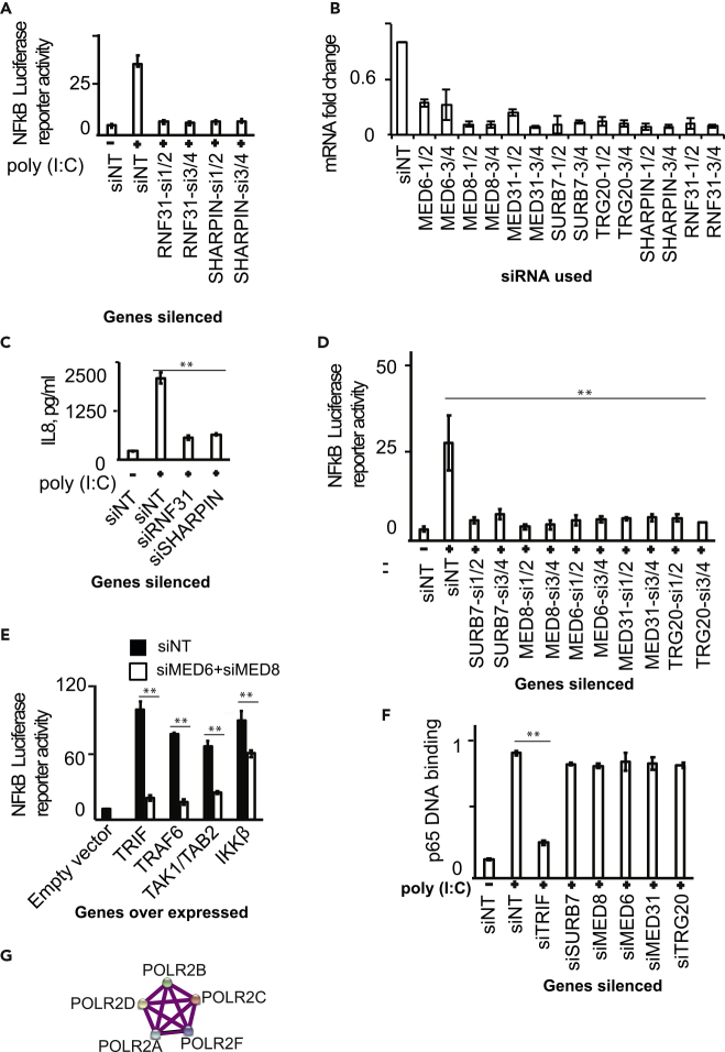
TLR3, a major innate immune pattern recognition receptor of RNA viruses, triggers inflammatory response through the transcription factor NF-κB. However, a genome-wide understanding of the genes and mechanisms regulating TLR3-mediated NF-κB activation is incomplete. We herein report the results of a human genome-wide RNAi screen that identified 591 proteins regulating TLR3-mediated NF-κB response. Bioinformatics analysis revealed several signaling modules including linear ubiquitination assembly complex and mediator protein complex network as regulators of TLR3 signaling. We further characterized the kinase ATM as a previously unknown positive regulator of TLR3 signaling. TLR3 pathway stimulation induced ATM phosphorylation and promoted interaction of ATM with TAK1, NEMO, IKKα, and IKKβ. Furthermore, ATM was determined to coordinate the assembly of NEMO with TAK1, IKKα, and IKKβ during TLR3 signaling. This study provided a comprehensive understanding of TLR3-mediated inflammatory signaling regulation and established a role for ATM in innate immune response.

Keywords: Biological Sciences; Cell Biology; Immunology.


Conflict of interest statement

Declaration of Interests Authors declare no competing interests.

Figures

None
Graphical abstract
Figure 1
Figure 1
RNAi Screening for Identifying TLR3-Mediated NF-κB Activation (A) Schematic showing RNAi screening methodology. Images correspond to the GFP signal in negative control siRNA and positive control siRNA targeting (si-TRIF). (B) GFP reporter assay efficiency for measuring NF-κB activation induced by poly(I:C) with and without nucleic acid sensing innate immune pathway genes. NF-κB-GFP reporter containing HEK293T cells were stimulated with poly(I:C) for 24 h, and images were quantified and expressed as percentage of GFP-positive cells. The values shown are mean ± SE of three independent experiments performed in triplicates. The statistical significance of the difference in values between groups was analyzed using unpaired two-tailed Student's t test, and p values <0.05 was considered statistically significant. ∗∗p value <0.01. Knockdown efficiencies of relevant siRNAs determined by western blot are shown in the inset; left and right panel of western blots, respectively, show scrambled siRNA or gene-specific siRNA-treated cells. (C–E) Enriched Molecular Function, Biological Processes, and Cellular Compartment categories among the hit genes obtained from RNAi screening, respectively. (F) Enrichment of disease categories among the screening hits. The plot was generated using Ingenuity Pathway Analysis software. GFP, green fluorescent protein. siNT, negative control siRNA.
Figure 2
Figure 2
Bioinformatics Analysis Identifies Gene Networks Regulating TLR3-Mediated NF-κB Activation (A) Interactome analysis using all hit genes revealed several known protein-protein associations. (B–D) Respectively shows signaling networks of linear ubiquitination complex, myosin, and mediator complex. The network was generated by STRING. The thickness of the connecting lines indicates the confidence level of known functional interactions between indicated proteins.
Figure 3
Figure 3
LUBAC and Mediator Complex Are Needed for TLR3 Signaling (A) Silencing of RNF31 and SHARPIN using two pairs of unique siRNAs reduced NF-κB luciferase reporter activation driven by poly(I:C) stimulation in HEK293T cells. (B) Efficiency of gene silencing is shown. Genes were targeted using pairs of siRNAs. The gene transcript levels were determined using qRT-PCR. The values correspond to mean ± SD of a triplicate experiment and are expressed as fold change of mRNA level relative to scrambled siNT sample (siNT value is taken as 1). The qRT-PCR results were computed through determination of relative Ct value, using the formula (Fold-change = 2(Ct of unstimulated – Ct of stimulated) Target gene/2(Ct of unstimulated – Ct of stimulated) Reference gene). (C) Silencing of RNF31 and SHARPIN reduced IL8 secretion driven by poly(I:C) stimulation in human primary monocytes. (D) Silencing of five component genes of mediator complex using two pairs of unique siRNAs reduced NF-κB luciferase reporter activation driven by poly(I:C) stimulation in HEK293T cells. (E) Mediator complex silencing did not affect the ability of HEK293T cells to support NF-κB luciferase reporter activation induced by ectopic expression of 50 ng each of TRIF, TRAF6, TAK1/TAB2, IKKβ for 24 h. (F) Mediator complex silencing did not affect the ability of p65 to bind to target DNA in poly(I:C) treated HEK293T cells. (G) A network of RNA polymerase II subunits uncovered in the current RNAi screening as hits. The values shown for (A and C–F) are mean ± SE of three independent experiments performed in triplicates. The statistical significance of the difference in values between groups was analyzed using an unpaired two-tailed Student's t test, and p values <0.05 were considered statistically significant. ∗∗p value <0.01.
Figure 4
Figure 4
ATM Kinase Is a Positive Regulator of TLR3-Mediated NF-κB Activation (A) Silencing of ATM using two pairs of unique siRNAs reduced NF-κB luciferase reporter activation driven by poly(I:C) stimulation in HEK293T cells. (B and C) Silencing of ATM reduced IL8 secretion driven by poly(I:C) stimulation, respectively, in HEK293T cells and human primary monocytes. (D) Type-I interferon beta reporter activity was unaffected by ATM silencing. (E) Poly(I:C) stimulation induced phosphorylation of ATM in HEK293T cells. The data are representative of at least three independent experiments. (F) ATM silencing reduced the ability of HEK293T cells to support NF-κB luciferase reporter activation induced by ectopic expression of 50 ng each of TRIF, TRAF6, TAK1/TAB2, IKKβ for 24 h. (G) ATM silencing reduced phosphorylation of Ikβ upon poly(I:C) stimulation. The data are representative of at least three independent experiments. (H) ATM silencing reduced the ability of p65 to bind to target DNA in poly(I:C)-treated HEK293T cells. (I) ATM silencing reduced NOD2-mediated NF-κB luciferase reporter activation driven by muramyl dipeptide (MDP) stimulation in HEK293T cells. The values shown for (A–F, H, and I) are mean ± SE of three independent experiments performed in triplicates. The statistical significance of the difference in values between groups was analyzed using an unpaired two-tailed Student's t test, and p values <0.05 were considered statistically significant. ∗∗p value <0.01, ∗p value <0.05. GAPDH, glyceraldehyde 3-phosphate dehydrogenase.
Figure 5
Figure 5
ATM Interacts with and Facilitates NEMO Signaling Complex Assembly (A) Endogenous ATM interacted with endogenous NEMO, TAK1, IKKα, and IKKβ upon stimulation of human primary monocytes with poly(I:C). (B) ATM expression was needed for NEMO to recruit TAK1, IKKα, and IKKβ upon stimulation of human primary monocytes with poly(I:C). ATM-silenced cells were stimulated with poly(I:C) for 120 min, and NEMO co-immunoprecipitation assay was performed. The data are representative of at least three independent experiments. GAPDH, glyceraldehyde 3-phosphate dehydrogenase; WCL, whole-cell lysate; IP, immunoprecipitation; IB, immunoblot.

References

    1. Ahmed S., Maratha A., Butt A.Q., Shevlin E., Miggin S.M. TRIF-mediated TLR3 and TLR4 signaling is negatively regulated by ADAM15. J. Immunol. 2013;190:2217–2228. - PubMed
    1. Alexopoulou L., Holt A.C., Medzhitov R., Flavell R.A. Recognition of double-stranded RNA and activation of NF-kappaB by Toll-like receptor 3. Nature. 2001;413:732–738. - PubMed
    1. Boisson B., Laplantine E., Prando C., Giliani S., Israelsson E., Xu Z., Abhyankar A., Israel L., Trevejo-Nunez G., Bogunovic D. Immunodeficiency, autoinflammation and amylopectinosis in humans with inherited HOIL-1 and LUBAC deficiency. Nat. Immunol. 2012;13:1178–1186. - PMC - PubMed
    1. Brencicova E., Diebold S.S. Nucleic acids and endosomal pattern recognition: how to tell friend from foe? Front. Cell Infect. Microbiol. 2013;3:37. - PMC - PubMed
    1. Brinkmann M.M., Spooner E., Hoebe K., Beutler B., Ploegh H.L., Kim Y.M. The interaction between the ER membrane protein UNC93B and TLR3, 7, and 9 is crucial for TLR signaling. J. Cell Biol. 2007;177:265–275. - PMC - PubMed