. 2020 Oct;586(7830):578-582.
doi: 10.1038/s41586-020-2608-y. Epub 2020 Jul 30.

ChAdOx1 nCoV-19 vaccine prevents SARS-CoV-2 pneumonia in rhesus macaques

Affiliations

ChAdOx1 nCoV-19 vaccine prevents SARS-CoV-2 pneumonia in rhesus macaques

Neeltje van Doremalen et al. Nature. 2020 Oct.

Erratum in

  • Publisher Correction: ChAdOx1 nCoV-19 vaccine prevents SARS-CoV-2 pneumonia in rhesus macaques.
    van Doremalen N, Lambe T, Spencer A, Belij-Rammerstorfer S, Purushotham JN, Port JR, Avanzato VA, Bushmaker T, Flaxman A, Ulaszewska M, Feldmann F, Allen ER, Sharpe H, Schulz J, Holbrook M, Okumura A, Meade-White K, Pérez-Pérez L, Edwards NJ, Wright D, Bissett C, Gilbride C, Williamson BN, Rosenke R, Long D, Ishwarbhai A, Kailath R, Rose L, Morris S, Powers C, Lovaglio J, Hanley PW, Scott D, Saturday G, de Wit E, Gilbert SC, Munster VJ. van Doremalen N, et al. Nature. 2021 Feb;590(7844):E24. doi: 10.1038/s41586-020-03099-2. Nature. 2021. PMID: 33469217 No abstract available.

Abstract

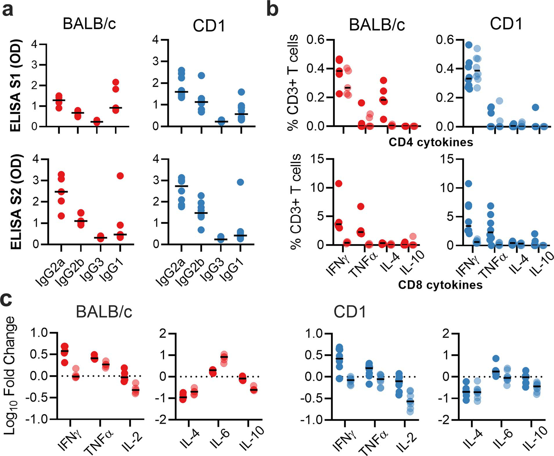
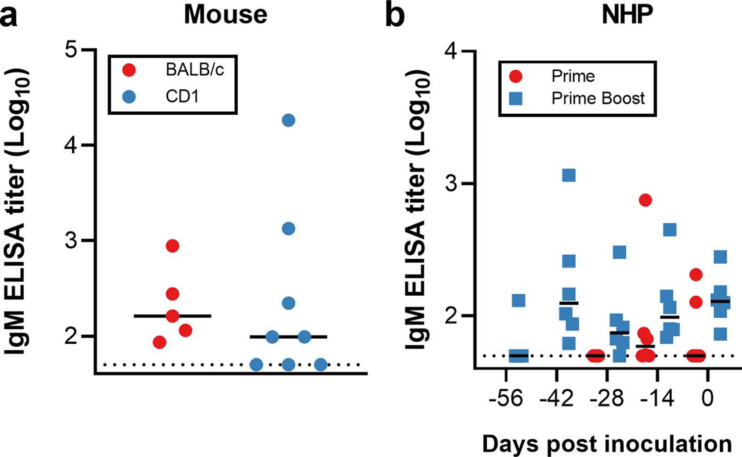
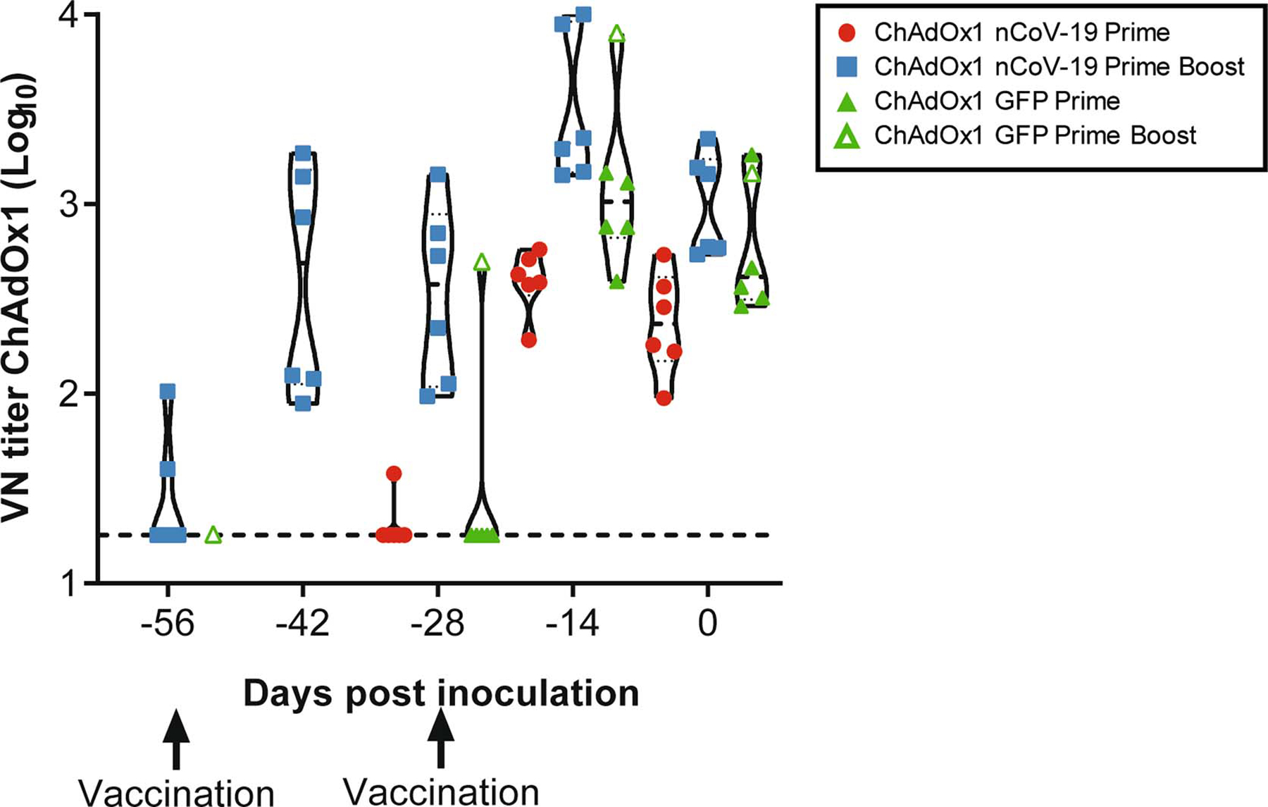
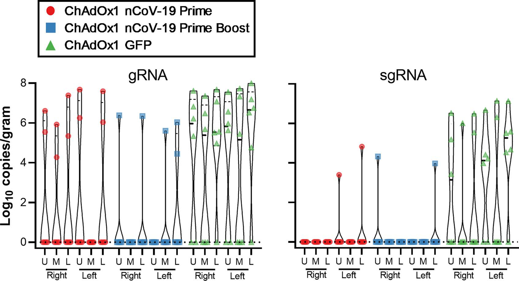
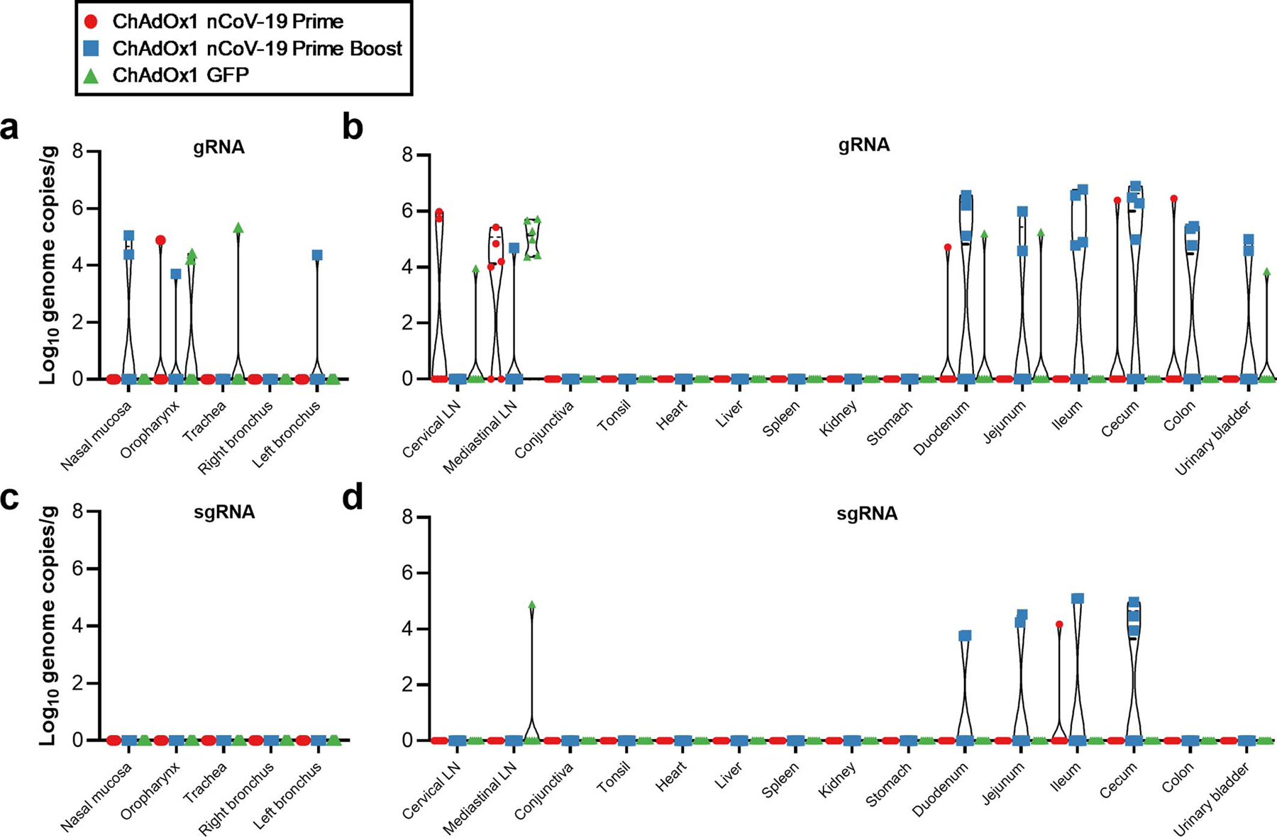
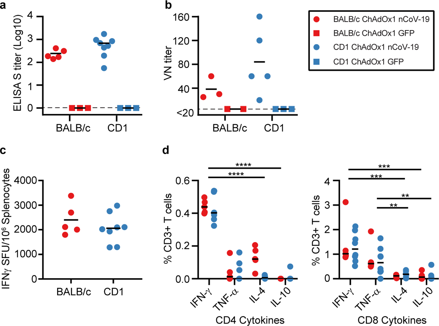
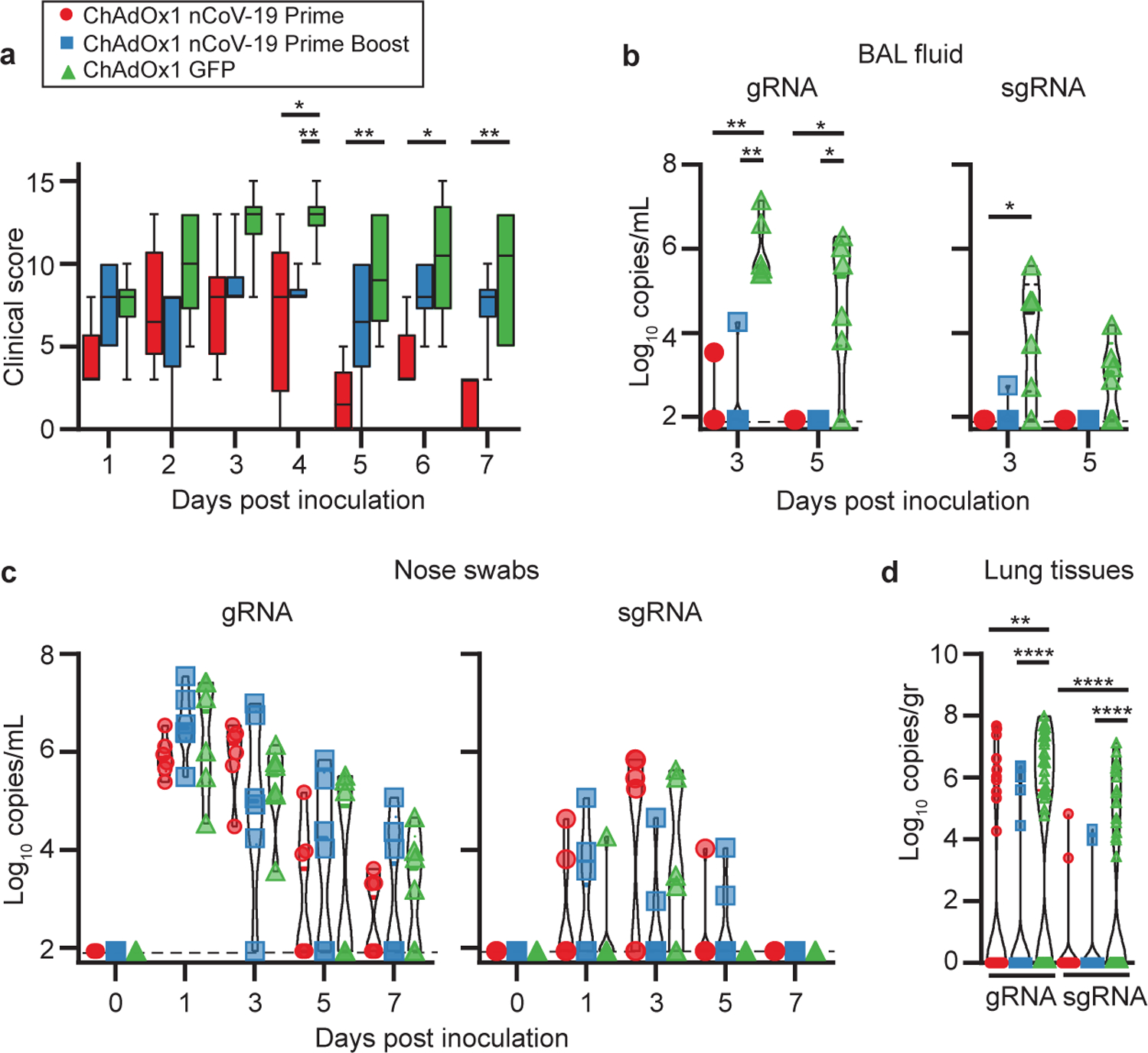
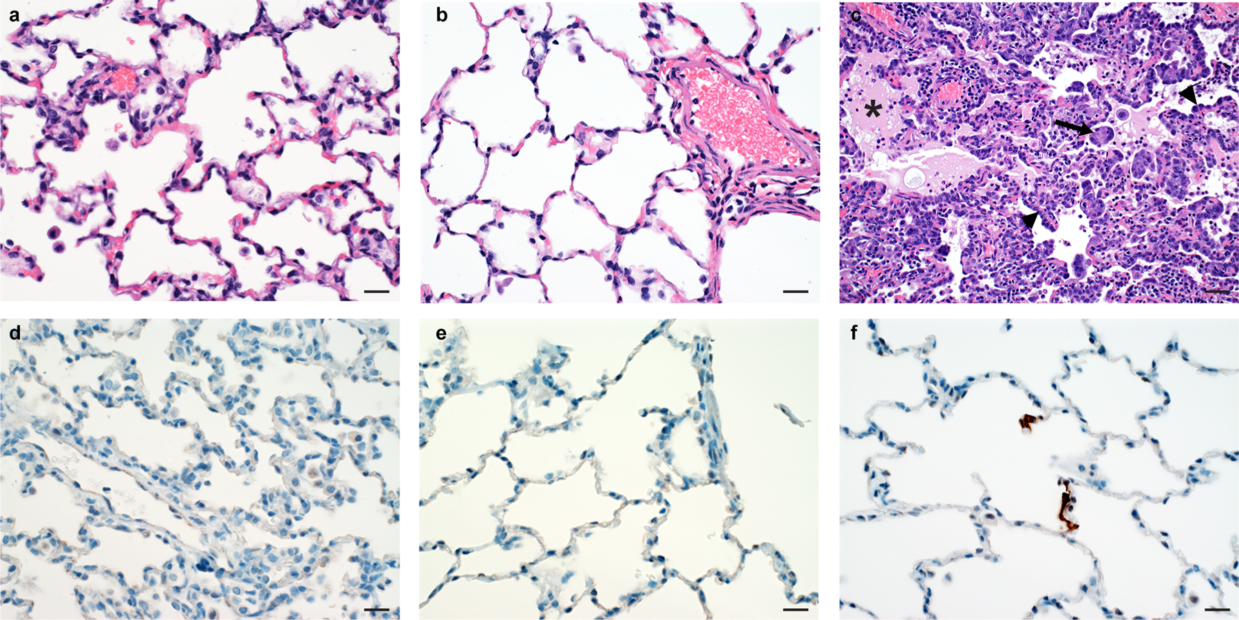
Severe acute respiratory syndrome coronavirus 2 (SARS-CoV-2) emerged in December 20191,2 and is responsible for the coronavirus disease 2019 (COVID-19) pandemic3. Vaccines are an essential countermeasure and are urgently needed to control the pandemic4. Here we show that the adenovirus-vector-based vaccine ChAdOx1 nCoV-19, which encodes the spike protein of SARS-CoV-2, is immunogenic in mice and elicites a robust humoral and cell-mediated response. This response was predominantly mediated by type-1 T helper cells, as demonstrated by the profiling of the IgG subclass and the expression of cytokines. Vaccination with ChAdOx1 nCoV-19 (using either a prime-only or a prime-boost regimen) induced a balanced humoral and cellular immune response of type-1 and type-2 T helper cells in rhesus macaques. We observed a significantly reduced viral load in the bronchoalveolar lavage fluid and lower respiratory tract tissue of vaccinated rhesus macaques that were challenged with SARS-CoV-2 compared with control animals, and no pneumonia was observed in vaccinated SARS-CoV-2-infected animals. However, there was no difference in nasal shedding between vaccinated and control SARS-CoV-2-infected macaques. Notably, we found no evidence of immune-enhanced disease after viral challenge in vaccinated SARS-CoV-2-infected animals. The safety, immunogenicity and efficacy profiles of ChAdOx1 nCoV-19 against symptomatic PCR-positive COVID-19 disease will now be assessed in randomized controlled clinical trials in humans.


Conflict of interest statement

Competing interests

SCG is a board member of Vaccitech and named as an inventor on a patent covering use of ChAdOx1-vectored vaccines and a patent application covering a SARS-CoV-2 (nCoV-19) vaccine (UK Patent Application No. 2003670.3). Teresa Lambe is named as an inventor on a patent application covering a SARS-CoV-2 (nCoV-19) vaccine (UK Patent Application No. 2003670.3). The University of Oxford and Vaccitech, having joint rights in the vaccine, entered into a partnership with AstraZeneca in April 2020 for further development, large-scale manufacture and global supply. Equitable access to the vaccine is a key component of the partnership. Neither Oxford University nor Vaccitech will receive any royalties during the pandemic period or from any sales of the vaccine in developing countries. The remaining authors declare no competing interests.

Figures

Extended Data Figure 1.
Extended Data Figure 1.. Antigen specific responses following ChAdOx1 nCov19 vaccination.
a. IgG subclass antibodies detected against S1 or S2 protein in sera of BALB/c or CD1 mice. b. Frequency of cytokine positive CD3+ T cells following stimulation of splenocytes with S1 pool (dark) or S2 pool (transparent) peptides in BALB/c (red) and CD1 (blue) mice. c. Log10 fold change in cytokine levels in supernatant from S1 (dark) and S2 (transparent) stimulated splenocytes when compared to corresponding unstimulated splenocyte sample for BALB/c and CD1 mice. n=5 (BALB/c) and 8 (CD1) animals examined over 1 independent experiment for all figure panels.
Extended Data Figure 2.
Extended Data Figure 2.. Spike-specific serum IgM.
a. Spike-specific serum IgM in mice 14 days post vaccination. n=5 (BALB/c) and 8 (CD1) animals examined over 1 independent experiment. b. Spike-specific serum IgM in NHPs upon prime-boost or prime-only vaccination. n=6 animals per group examined over 2 independent experiments.
Extended Data Figure 3.
ChAdOx1 neutralizing antibodies in serum of vaccinated NHPs. Control animal with prime-boost regimen is highlighted with open triangle symbol. VN = virus neutralizing. n=6 animals per group examined over 2 independent experiments.
Extended Data Figure 4.
Extended Data Figure 4.. Serum cytokines in rhesus macaques challenged with SARS-CoV-2.
Fold increase in cytokines in serum compared to pre-challenge values. ** = p-value<0.01; Line = median. Statistical significance determined via two-tailed Mann-Whitney test. P-values: IFN-γ = 0.0087; IL-10 = 0.0043; IL-13 = 0.0043 and 0.0065. n=6 animals per group examined over 2 independent experiments.
Extended Data Figure 5.
Viral load in lung tissue of rhesus macaques challenged with SARS-CoV-2 at 7 DPI.
Extended Data Figure 6.
Extended Data Figure 6.. Viral load in tissues of rhesus macaques challenged with SARS-CoV-2 at 7 DPI.
a. Viral gRNA in respiratory tissues excluding lung tissue. b. Viral gRNA in non-respiratory tissues. c. Viral sgRNA in respiratory tissues excluding lung tissue. d. Viral sgRNA in non-respiratory tissues.
Figure 1:
Figure 1:. Humoral and cellular immune responses to ChAdOx1 nCoV-19 vaccination in mice.
a. End point titer of serum IgG detected against S protein at 14 days post vaccination. No positive responses were detected in the control group. n=5, 3, 8, and 3 animals respectively examined over 1 independent experiment. b. Virus neutralizing titer in serum at 9 days post vaccination. n=3, 2, 5, and 3 animals respectively examined over 1 independent experiment. c. Summed IFN-γ ELISpot responses in splenocytes toward peptides spanning the spike protein at 14 days post vaccination. Control mice had low (<100 SFU) responses. n=5 and 8 animals respectively examined over 1 independent experiment. d. Summed frequency of spike-specific cytokine positive CD3+ T cells at 14 days post vaccination. P-value left panel: <0.0001. P-value right panel 0.0002 (IFN-γ−IL-4); 0.0001 (IFN-γ-IL-10); 0.0054 (TNF-α-IL-4); 0.0022 (TNF-α-IL-10). n=5 and 8 animals respectively examined over 1 independent experiment. BALB/c = red; CD1 = blue; vaccinated = circle; control = square; dotted line = limit of detection; line = mean; SFU = spot-forming units; Spl. = splenocytes; * = p<0.05. Statistical significance determined via 2-way ANOVA (repeated measure) and post-hoc positive test.
Figure 2.
Figure 2.. Humoral and cellular immune responses to ChAdOx1 nCoV-19 vaccination in rhesus macaques.
a. Study schedule for NHPs. V = vaccination; E = exam; C = exam and challenge; N = exam and necropsy. Violin plots of b. endpoint IgG titer in serum against trimeric spike protein, c. VN titer in serum and d. Summed IFN-γ ELISpot responses in PBMCs collected at 0 DPI toward peptides spanning the spike protein. Red circles = prime-only vaccine; blue squares = prime-boost vaccine; green triangle = controls; dotted line = limit of detection; SFU = spot-forming units; VN= virus-neutralizing; * = p-value=0.0313. Statistical significance determined via two-tailed Wilcoxon test.
Figure 3.
Figure 3.. Clinical signs and viral load in rhesus macaques inoculated with SARS-CoV-2 after vaccination with ChAdOx1 nCoV-19.
a. Boxplot of 25th to 75th percentile with median as centre and whiskers of 5th to 95th percentile clinical score in NHPs. n=6 animals per group examined over 2 independent experiments. P-values = 0.0455 (Prime-control D3); 0.0238 (Prime-control D4); 0.0043 (Prime-boost-control D4); 0.0043 (Prime-control D5); 0.0152 (Prime-control D6); 0.0022 (Prime-control D7). Violin plot of viral load in b. BAL fluid (* = p-value 0.0152; ** = p-value 0.0022) and c. nose swabs obtained from rhesus macaques. d. Violin plot of viral load in lung tissue. n=6 lung lobes of 6 animals per group examined over 2 independent experiments. **=p-value=0.0011; ****=p-value<0.0001. Dotted line = limit of detection. Statistical significance determined via two-tailed Mann-Whitney test.
Figure 4.
Figure 4.. Histological changes in lungs of rhesus macaques on 7 DPI.
No histological changes were observed in the lungs of ChAdOx1 nCoV-19 prime (a) and prime-boost (b) vaccinated animals. c) Interstitial pneumonia with edema (asterisk), type II pneumocyte hyperplasia (arrowhead) and syncytial cells (arrow) in control animals. No SARS-CoV-2 antigen was detected by immunohistochemistry in the lungs of ChAdOx1 nCoV-19 prime (d) and prime-boost (e) vaccinated animals. f) SARS-CoV-2 antigen (visible as red-brown staining) was detected by immunohistochemistry in type I and type II pneumocytes in the lungs of control animals. 18 sections, taken from 6 different lung lobes are evaluated for each animal; a representative lesion from each group was selected for the figure. Magnification: 400x, scale bar = 20 µm.

Update of

  • ChAdOx1 nCoV-19 vaccination prevents SARS-CoV-2 pneumonia in rhesus macaques.
    van Doremalen N, Lambe T, Spencer A, Belij-Rammerstorfer S, Purushotham JN, Port JR, Avanzato V, Bushmaker T, Flaxman A, Ulaszewska M, Feldmann F, Allen ER, Sharpe H, Schulz J, Holbrook M, Okumura A, Meade-White K, Pérez-Pérez L, Bissett C, Gilbride C, Williamson BN, Rosenke R, Long D, Ishwarbhai A, Kailath R, Rose L, Morris S, Powers C, Lovaglio J, Hanley PW, Scott D, Saturday G, de Wit E, Gilbert SC, Munster VJ. van Doremalen N, et al. bioRxiv [Preprint]. 2020 May 13:2020.05.13.093195. doi: 10.1101/2020.05.13.093195. bioRxiv. 2020. Update in: Nature. 2020 Oct;586(7830):578-582. doi: 10.1038/s41586-020-2608-y. PMID: 32511340 Free PMC article. Updated. Preprint.

References

    1. Zhu N et al. A Novel Coronavirus from Patients with Pneumonia in China, 2019. N Engl J Med 382, 727–733, doi: 10.1056/NEJMoa2001017 (2020). - DOI - PMC - PubMed
    1. Wu F et al. A new coronavirus associated with human respiratory disease in China. Nature 579, 265–269, doi: 10.1038/s41586-020-2008-3 (2020). - DOI - PMC - PubMed
    1. WHO. Coronavirus disease (COVID-19) Situation Report 113, <https://www.who.int/docs/default-source/coronaviruse/situation-reports/2...> (2020).
    1. Lurie N, Saville M, Hatchett R & Halton J Developing Covid-19 Vaccines at Pandemic Speed. N Engl J Med, doi: 10.1056/NEJMp2005630 (2020). - DOI - PubMed
    1. Dicks MD et al. A novel chimpanzee adenovirus vector with low human seroprevalence: improved systems for vector derivation and comparative immunogenicity. PLoS One 7, e40385, doi: 10.1371/journal.pone.0040385 (2012). - DOI - PMC - PubMed

References Methods

    1. Yasui F et al. Prior immunization with severe acute respiratory syndrome (SARS)-associated coronavirus (SARS-CoV) nucleocapsid protein causes severe pneumonia in mice infected with SARS-CoV. J Immunol 181, 6337–6348, doi: 10.4049/jimmunol.181.9.6337 (2008). - DOI - PubMed
    1. Bewig B & Schmidt WE Accelerated titering of adenoviruses. Biotechniques 28, 870–873, doi: 10.2144/00285bm08 (2000). - DOI - PubMed
    1. Maizel JV Jr., White DO & Scharff MD The polypeptides of adenovirus. I. Evidence for multiple protein components in the virion and a comparison of types 2, 7A, and 12. Virology 36, 115–125, doi: 10.1016/0042-6822(68)90121-9 (1968). - DOI - PubMed
    1. Corman VM et al. Detection of 2019 novel coronavirus (2019-nCoV) by real-time RT-PCR. Euro Surveill 25, doi: 10.2807/1560-7917.ES.2020.25.3.2000045 (2020). - DOI - PMC - PubMed
    1. Wolfel R et al. Virological assessment of hospitalized patients with COVID-2019. Nature, doi: 10.1038/s41586-020-2196-x (2020). - DOI - PubMed

Publication types

MeSH terms