

Matrix Stiffness Modulates Patient-Derived Glioblastoma Cell Fates in Three-Dimensional Hydrogels
- PMID: 32731804
- PMCID: PMC7984937
- DOI: 10.1089/ten.TEA.2020.0110
Matrix Stiffness Modulates Patient-Derived Glioblastoma Cell Fates in Three-Dimensional Hydrogels
Abstract
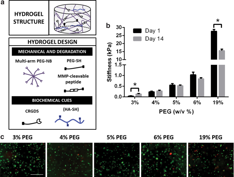
Cancer progression is known to be accompanied by changes in tissue stiffness. Previous studies have primarily employed immortalized cell lines and 2D hydrogel substrates, which do not recapitulate the 3D tumor niche. How matrix stiffness affects patient-derived cancer cell fate in 3D remains unclear. In this study, we report a matrix metalloproteinase-degradable poly(ethylene-glycol)-based hydrogel platform with brain-mimicking biochemical cues and tunable stiffness (40-26,600 Pa) for 3D culture of patient-derived glioblastoma xenograft (PDTX GBM) cells. Our results demonstrate that decreasing hydrogel stiffness enhanced PDTX GBM cell proliferation, and hydrogels with stiffness 240 Pa and below supported robust PDTX GBM cell spreading in 3D. PDTX GBM cells encapsulated in hydrogels demonstrated higher drug resistance than 2D control, and increasing hydrogel stiffness further enhanced drug resistance. Such 3D hydrogel platforms may provide a valuable tool for mechanistic studies of the role of niche cues in modulating cancer progression for different cancer types.
Keywords: glioblastoma; hydrogels; in vitro cancer models; patient-derived cells; stiffness.
Conflict of interest statement
No competing financial interests exist.
Figures


Similar articles
-
Gradient hydrogels for screening stiffness effects on patient-derived glioblastoma xenograft cellfates in 3D.J Biomed Mater Res A. 2021 Jun;109(6):1027-1035. doi: 10.1002/jbm.a.37093. Epub 2020 Sep 21. J Biomed Mater Res A. 2021. PMID: 32862485
-
Bioengineered 3D brain tumor model to elucidate the effects of matrix stiffness on glioblastoma cell behavior using PEG-based hydrogels.Mol Pharm. 2014 Jul 7;11(7):2115-25. doi: 10.1021/mp5000828. Epub 2014 Apr 29. Mol Pharm. 2014. PMID: 24712441
-
Dynamically Crosslinked Poly(ethylene-glycol) Hydrogels Reveal a Critical Role of Viscoelasticity in Modulating Glioblastoma Fates and Drug Responses in 3D.Adv Healthc Mater. 2023 Jan;12(1):e2202147. doi: 10.1002/adhm.202202147. Epub 2022 Nov 16. Adv Healthc Mater. 2023. PMID: 36239185 Free PMC article.
-
How Hydrogel Stiffness Affects Adipogenic Differentiation of Mesenchymal Stem Cells under Controlled Morphology.ACS Appl Bio Mater. 2023 Sep 18;6(9):3441-3450. doi: 10.1021/acsabm.3c00159. Epub 2023 Apr 16. ACS Appl Bio Mater. 2023. PMID: 37061939 Review.
-
Glioblastoma mechanobiology at multiple length scales.Biomater Adv. 2024 Jun;160:213860. doi: 10.1016/j.bioadv.2024.213860. Epub 2024 Apr 15. Biomater Adv. 2024. PMID: 38640876 Review.
Cited by
-
Biofabrication of Modular Spheroids as Tumor-Scale Microenvironments for Drug Screening.Adv Healthc Mater. 2023 Jun;12(14):e2201581. doi: 10.1002/adhm.202201581. Epub 2022 Dec 25. Adv Healthc Mater. 2023. PMID: 36495232 Free PMC article.
-
Different Decellularization Methods in Bovine Lung Tissue Reveals Distinct Biochemical Composition, Stiffness, and Viscoelasticity in Reconstituted Hydrogels.ACS Appl Bio Mater. 2023 Feb 20;6(2):793-805. doi: 10.1021/acsabm.2c00968. Epub 2023 Feb 2. ACS Appl Bio Mater. 2023. PMID: 36728815 Free PMC article.
-
The multifaceted mechanisms of malignant glioblastoma progression and clinical implications.Cancer Metastasis Rev. 2022 Dec;41(4):871-898. doi: 10.1007/s10555-022-10051-5. Epub 2022 Aug 3. Cancer Metastasis Rev. 2022. PMID: 35920986 Free PMC article. Review.
-
Enhanced anti-tumor efficacy of "IL-15 and CCL19" -secreting CAR-T cells in human glioblastoma orthotopic xenograft model.Front Oncol. 2025 Mar 19;15:1539055. doi: 10.3389/fonc.2025.1539055. eCollection 2025. Front Oncol. 2025. PMID: 40177238 Free PMC article.
-
Application of Nano-Inspired Scaffolds-Based Biopolymer Hydrogel for Bone and Periodontal Tissue Regeneration.Polymers (Basel). 2022 Sep 10;14(18):3791. doi: 10.3390/polym14183791. Polymers (Basel). 2022. PMID: 36145936 Free PMC article. Review.
References
-
- Wells, R.G. The role of matrix stiffness in regulating cell behavior. Hepatology 47, 1394, 2008 - PubMed
-
- Yeung, T., Georges, P.C., Flanagan, L.A., et al. . Effects of substrate stiffness on cell morphology, cytoskeletal structure, and adhesion. Cell Motil Cytoskeleton 60, 24, 2005 - PubMed
-
- Engler, A.J., Sen, S., Sweeney, H.L., and Discher, D.E.. Matrix elasticity directs stem cell lineage specification. Cell 126, 677, 2006 - PubMed
Publication types
MeSH terms
Substances
Grants and funding
LinkOut - more resources
Full Text Sources
Other Literature Sources
Medical
