. 2020 Jul 31;369(6503):554-557.
doi: 10.1126/science.abb3758.

In-cell architecture of an actively transcribing-translating expressome

Affiliations

In-cell architecture of an actively transcribing-translating expressome

Francis J O'Reilly et al. Science. .

Abstract

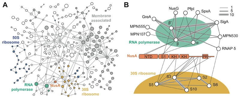
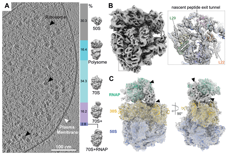
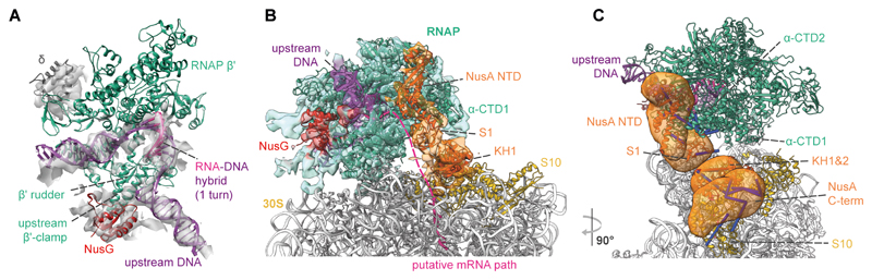
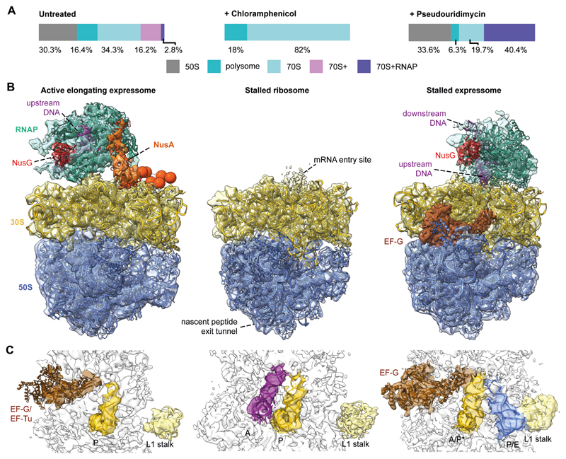
Structural biology studies performed inside cells can capture molecular machines in action within their native context. In this work, we developed an integrative in-cell structural approach using the genome-reduced human pathogen Mycoplasma pneumoniae We combined whole-cell cross-linking mass spectrometry, cellular cryo-electron tomography, and integrative modeling to determine an in-cell architecture of a transcribing and translating expressome at subnanometer resolution. The expressome comprises RNA polymerase (RNAP), the ribosome, and the transcription elongation factors NusG and NusA. We pinpointed NusA at the interface between a NusG-bound elongating RNAP and the ribosome and propose that it can mediate transcription-translation coupling. Translation inhibition dissociated the expressome, whereas transcription inhibition stalled and rearranged it. Thus, the active expressome architecture requires both translation and transcription elongation within the cell.


Conflict of interest statement

Competing interests: The authors have no competing interests.

Figures

Fig. 1
Fig. 1. Crosslink-based protein interaction map of M. pneumoniae proteome.
(A) 577 distinct PPIs identified at 5% PPI-level FDR (interactions to 8 abundant glycolytic enzymes and chaperones are removed for clarity). Membrane-associated proteins are shown in grey. Circle diameter indicates relative protein size. Blue: 50S ribosomal proteins; yellow: 30S ribosomal proteins; green: RNAP; orange: NusA. Each edge represents one or more crosslinks. (B) Interactors of RNAP and NusA. NusA NTD, S1, KH domains, and proline rich region (PR) are annotated. Line thickness represents the number of identified crosslinks.
Fig. 2
Fig. 2. In-cell cryo-ET reveals the presence of an RNAP-ribosome supercomplex.
(A) Left: tomographic slice of a M. pneumoniae cell. Right: classification of 108,501 ribosome sub-tomograms from M. pneumoniae cells. (B) Left: 5.6 Å in-cell 70S ribosome density. Insert: density near the peptide exit tunnel (dashed circle) shows two helices not accounted for by the fitted homology model (L22 and L29). (C) 9.2 Å in-cell structure of RNAP-ribosome supercomplexes (2.8% in (A)), fitted with homology models. Arrowheads indicate remaining unassigned density.
Fig. 3
Fig. 3. Integrative model of the M. pneumoniae elongating expressome.
(A) Cryo-EM density of RNAP corresponding to the DNA, RNA-DNA hybrid, upstream β’ clamp, δ subunit and NusG, accommodates one turn of the RNA-DNA hybrid consistent with an elongating RNAP. (B) Integrative model with cryo-EM density of the RNAP-NusG-NusA-ribosome elongating expressome. Structured regions are represented as colored cartoons. The electron density colors represent the subunits occupying the corresponding volumes. Coarse-grained regions are not shown. Schematic of the putative mRNA path refers to the shortest distance between mRNA exit and entry sites. (C) The mRNA exit tunnel face of RNAP is covered by NusA. Localization probability densities for NusA domains are shown in orange. Crosslinks between NusA and other proteins are shown. Satisfied crosslinks (<35 Å) are in blue, overlong crosslinks are in red.
Fig. 4
Fig. 4. Stalling translation or transcription alters the expressome architecture in cells.
(A) Classification of sub-tomograms in untreated, Cm- and PUM-treated M. pneumoniae cells revealed shifts in ribosome populations following perturbations. (B) Models of RNAP-ribosome supercomplexes and the Cm-stalled ribosome. Left: in untreated cells, the expressome compromises an actively elongating RNAP and ribosome. Center: Cm decoupled the ribosome and RNAP. Right: in PUM-treated cells, the ribosome encounters the stalled RNAP. (C) Ribosome tRNA occupancy states. In untreated cells, densities for P-site tRNA and elongation factors densities were visible, indicating a translating ribosome. Upon addition of Cm, A and P site tRNAs were observed indicating a stalled ribosome. In PUM-treated cells, presence of EF-G and hybrid A/P* and P/E site tRNAs suggested a pre-translocation stalled state.

Comment in

References

    1. Santangelo TJ, Artsimovitch I. Termination and antitermination: RNA polymerase runs a stop sign. Nat Rev Microbiol. 2011;9:319–329. - PMC - PubMed
    1. McGary K, Nudler E. RNA polymerase and the ribosome: the close relationship. Curr Opin Microbiol. 2013;16:112–117. - PMC - PubMed
    1. Conn AB, Diggs S, Tam TK, Blaha GM. Two Old Dogs, One New Trick: A Review of RNA Polymerase and Ribosome Interactions during Transcription-Translation Coupling. Int J Mol Sci. 2019;20:E2595. - PMC - PubMed
    1. Landick R, Carey J, Yanofsky C. Translation activates the paused transcription complex and restores transcription of the trp operon leader region. Proc Natl Acad Sci U S A. 1985;82:4663–4667. - PMC - PubMed
    1. Proshkin S, Rahmouni AR, Mironov A, Nudler E. Cooperation between translating ribosomes and RNA polymerase in transcription elongation. Science. 2010;328:504–508. - PMC - PubMed

Publication types

MeSH terms

Substances