. 2020 Jul 30;11(1):3794.
doi: 10.1038/s41467-020-17578-7.

Paraventricular hypothalamus mediates diurnal rhythm of metabolism

Affiliations

Paraventricular hypothalamus mediates diurnal rhythm of metabolism

Eun Ran Kim et al. Nat Commun. .

Abstract

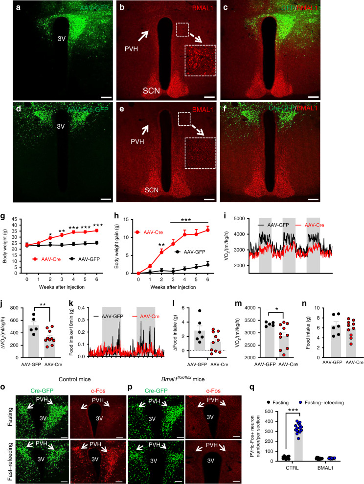
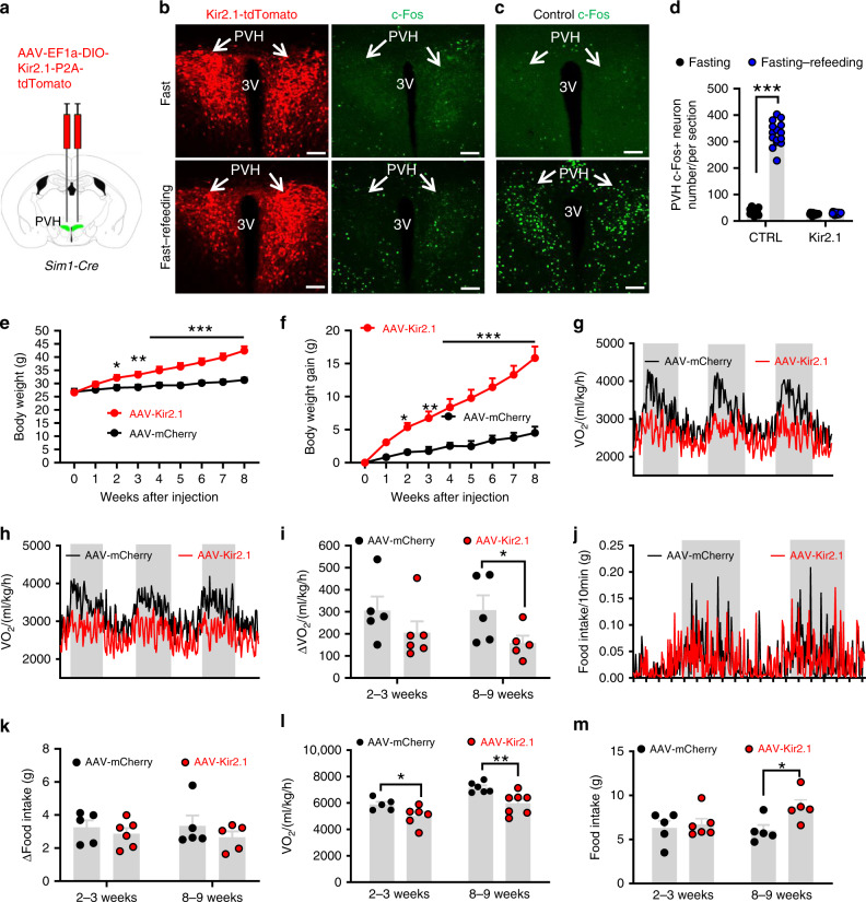
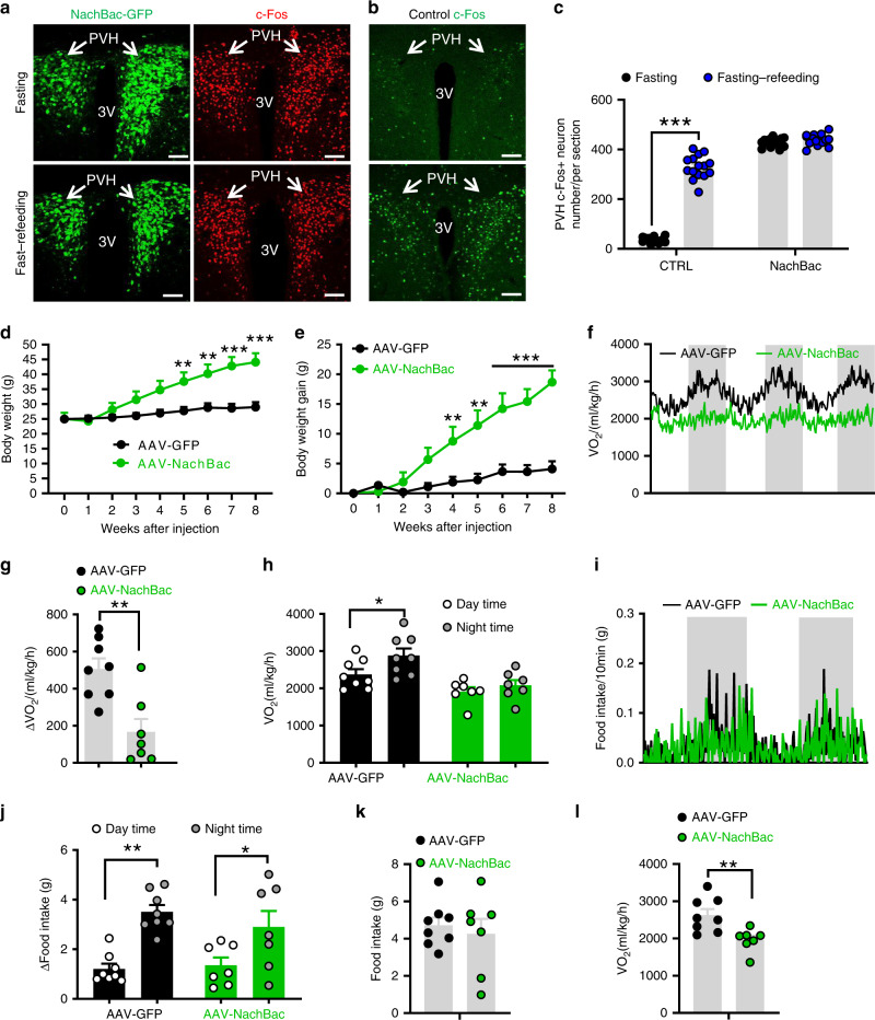
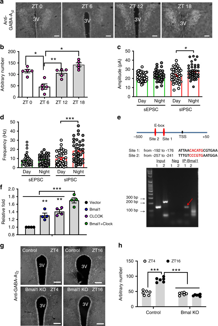
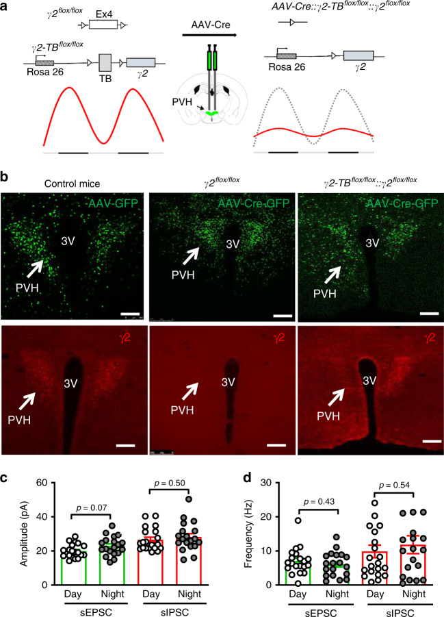
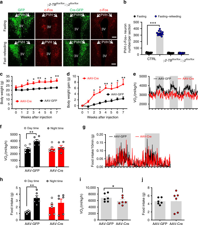
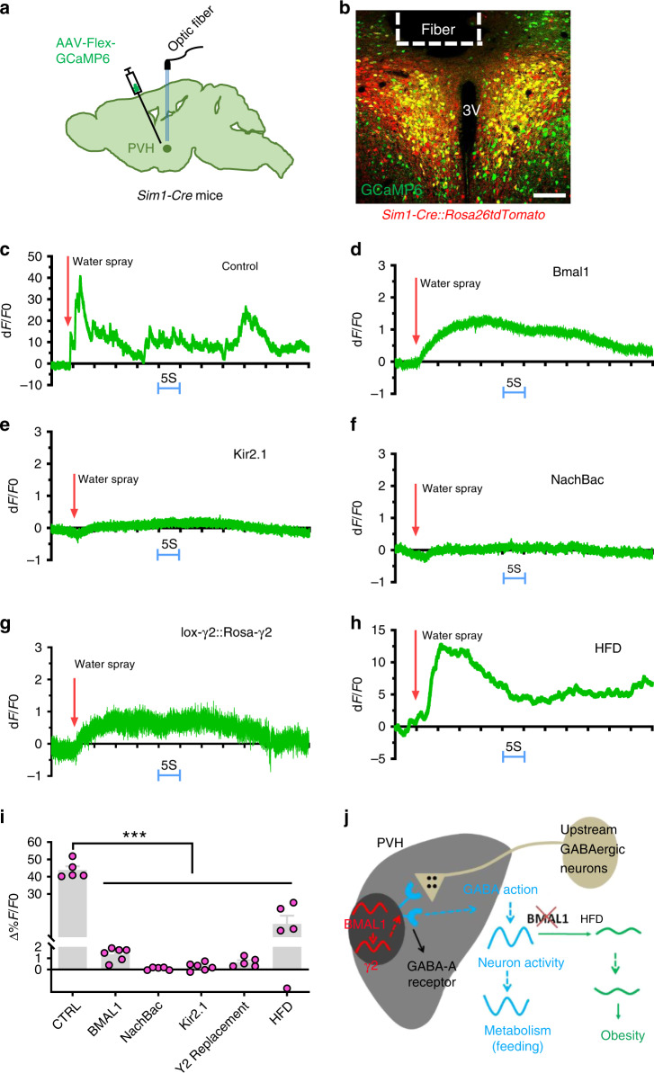
Defective rhythmic metabolism is associated with high-fat high-caloric diet (HFD) feeding, ageing and obesity; however, the neural basis underlying HFD effects on diurnal metabolism remains elusive. Here we show that deletion of BMAL1, a core clock gene, in paraventricular hypothalamic (PVH) neurons reduces diurnal rhythmicity in metabolism, causes obesity and diminishes PVH neuron activation in response to fast-refeeding. Animal models mimicking deficiency in PVH neuron responsiveness, achieved through clamping PVH neuron activity at high or low levels, both show obesity and reduced diurnal rhythmicity in metabolism. Interestingly, the PVH exhibits BMAL1-controlled rhythmic expression of GABA-A receptor γ2 subunit, and dampening rhythmicity of GABAergic input to the PVH reduces diurnal rhythmicity in metabolism and causes obesity. Finally, BMAL1 deletion blunts PVH neuron responses to external stressors, an effect mimicked by HFD feeding. Thus, BMAL1-driven PVH neuron responsiveness in dynamic activity changes involving rhythmic GABAergic neurotransmission mediates diurnal rhythmicity in metabolism and is implicated in diet-induced obesity.


Conflict of interest statement

The authors declare no competing interests.

Figures

Fig. 1
Fig. 1. Adult deletion of PVH BMAL1 disrupted diurnal metabolism and caused obesity.
a–n Bmal1flox/flox mice (8–10 weeks old) received bilateral injections of AAV-GFP or AAV-Cre-GFP and were used for immunostaining BMAL1 expression and body weight studies. ac Brain sections from AAV-GFP-injected mice were examined for GFP expression (a), BMAL1 (b), and merged (c). df Brain section from AAV-Cre-GFP-injected mice were examined for GFP (d), BMAL1 (e), and their merged expression (f). Arrows pointing to BMAL1 expression in GFP (b) and Cre-injected mice (e). Insets in b and e showing BMAL1 expression in a higher magnification. PVH paraventricular hypothalamus, 3V the third ventricle, SCN superachiasmatic nucleus. Scale bar = 200 µM. gh Weekly body weight (g, two-way ANOVA, n = 5 for GFP and 10 for Cre, F(6, 84) = 3.382, ***p < 0.0001, body weight at 6 weeks between GFP vs Cre) and net increases in body weight (h, two-way ANOVA, n = 5 for GFP and 10 for Cre, F(6, 84) = 7.051, ***p < 0.0001, body weight at 6 weeks between GFP vs Cre) were followed. in Real time traces showing energy expenditure (i) and feeding (k) measured by CLAMS 3 weeks after viral delivery, comparison in difference between day and night periods of energy expenditure (j, unpaired two-tailed Student’s t-test, n = 5 for GFP and 10 for Cre, t = 3.014, d.f. = 13, p = 0.01) and feeding (l), and comparison in daily feeding (m, unpaired two-tailed Student’s t-test, n = 5 for GFP and 10 for Cre, t = 2.168, d.f. = 13, *p = 0.0493) and energy expenditure (n) between groups. oq Control or Bmalflox/flox mice with AAV-Cre-GFP injections were either overnight fasted alone or overnight fasted with 2 h refeeding, and then immunostained for c-Fos. Representative expression of GFP and c-Fos in the PVH in GFP (o) and BMAL1 deleted mice (p). At least three mice with five sections each containing the PVH were used for counting the number of c-Fos in the PVH. Arrows point to the PVH proper; 3V the third ventricle. q Comparison in average number of c-Fos-positive neurons in the PVH (two-way ANOVA, n = 15/each, F(1, 56) = 536.8, ***p < 0.0001 for GFP fasting vs fasting–refeeding, p = 0.9918 for Cre fasting vs fasting–refeeding). All data presented at mean ± SEM. Source data are provided as a Source Data file.
Fig. 2
Fig. 2. Clamping PVH neuron activity at a low lever disrupted diurnal metabolism and caused obesity.
Sim1-Cre mice (8–10 weeks old) received injections of AAV-FLEX-mCherry or AAV-DIO-EF1a-Kir2.1-P2A-dTomato vectors to bilateral PVH and used for studies. a Diagram showing injections of viral vectors to the PVH of Sim1-Cre mice. bd Expression of dTomato in the PVH after the Kir2.1 virus expression (b, left panels), and representative expression of c-Fos expression in the PVH after overnight fasting alone (top panels) or overnight fast with 2 h refeeding (bottom panels) in Kir2.1 mice (b, right panels) and control (c). At least three mice with five sections containing the PVH were used for counting the number of c-Fos in the PVH. Arrows point to the PVH proper. d Comparison of average number of c-Fos positive neurons in the PVH (two-way ANOVA, n = 15 each, F(1, 56) = 544.4, ***p < 0.0001, control fasting vs fasting–refeeding; and p = 0.9909 Kir2.1 fasting vs fasting–refeeding). em Weekly body weight (e, two-way ANOVA, n = 5 for control and 6 for Kir2.1, F(8, 171) = 4.022, ***p < 0.0001, body weight at 8-9 weeks after injection between Control and Kir2.1) and weekly net body gain (f, two-way ANOVA, n = 5 for control and 6 for Kir2.1, F(8, 171) = 6.401, ***p < 0.0001, body weight at 8-9 weeks after injection between Control and Kir2.1) were followed, and real time traces showing energy expenditure measurements at 2–3 weeks (g) and 8–9 weeks (h), and feeding 2–3 weeks (j) after viral delivery; comparison in the difference of energy expenditure (i, two-way ANOVA, n = 5 for Control and 6 for Kir2.1, F(1, 17) = 1.138, *p = 0.0378, 8-9 weeks after injection between Control and Kir2.1) and feeding (k) between day and night both 2–3 and 8–9 weeks after viral delivery; daily energy expenditure (l, two-way ANOVA, n = 5 for Control and 6 for Kir2.1, F(1, 20) = 25.99, *p = 0.026 2–3 weeks after injection and **p = 0.0059 for 8–9 weeks after injection between Control and Kir2.1) and feeding (m, two-way ANOVA, n = 5 for Control and 6 for Kir2.1, F(1, 20) = 3.292, *p < 0.0399, 8-9 weeks after injection control vs Kir2.1) both 2–3 and 8–9 weeks after viral delivery were shown. All data presented at mean ± SEM. PVH paraventricular hypothalamus, 3V the third ventricle. Scale bar = 200 µM. Source data are provided as a Source Data file.
Fig. 3
Fig. 3. Clamping PVH neuron activity at a high lever disrupted diurnal metabolism and caused obesity.
Sim1-Cre mice (8–10 weeks old) received injections of AAV-FLEX-GFP or AAV-EF1a-FLEX-EGFP-P2A-mNachBac vectors to bilateral PVH and used for studies. a, b Expression of EGFP in the PVH after the NachBac virus expression (a, left panels), and representative expression of c-Fos expression in the PVH after overnight fast alone (top panels) or overnight fast with 2 h refeeding (bottom panels) in NachBac mice (a, right panels) and control (b). c Comparison of number of neurons with c-Fos expression in the PVH between fast and fast–refeeding in control and NachBac mice (two-way ANOVA, n = 15/each, F(1, 56) = 385.3, ***p < 0.0001 Control fasting vs refeeding and p = 0.7212 NachBac fasting vs refeeding). At least three mice with five sections containing the PVH were used for counting the number of c-Fos in the PVH. Arrows point to the PVH proper. dh Weekly body weight (d, two-way ANOVA, n = 8 for GFP and 7 for NachBac, F(8, 117) = 4.252, ***p < 0.0001, body weight at 8 weeks after injection between Control and NachBac), weekly net body gain (e, two-way ANOVA, n = 8 for GFP and 7 for NachBac, F(8, 171) = 7.256, ***p < 0.0001, body weight at 8 weeks after injection between Control and NachBac) were followed, real time traces showing energy expenditure (f), comparison in the difference of energy expenditure between day and night (g, unpaired two-tailed Student’s t-test, n = 8 for GFP and 7 for NachBac, t = 3.836, d.f. = 13, **p = 0.0021), and differences between day and night periods within groups (h, two-way ANOVA, n = 8 for GFP and 7 for NachBac, F(3, 26) = 9.314, *p = 0.0488 control GFP day vs night and p = 0.8657, NachBac day vs night). i, j Real time traces showing feeding (i) and comparison in the difference of feeding between day and night (j, two-way ANOVA, n = 8 for GFP and 7 for NachBac, F(3, 26) = 9.257, ***p = 0.0008 Control GFP day vs night, and *p = 0.0458 NachBac day vs night). jl Daily feeding (k) and energy expenditure (l, unpaired two-tailed Student’s t-test, n = 8 for GFP and 7 for NachBac, t = 3.240, d.f. = 13, **p = 0.0064) were compared between groups. PVH paraventricular hypothalamus, 3V the third ventricle. Scale bar = 200 µM. All data presented as mean ± SEM. Source data are provided as a Source Data file.
Fig. 4
Fig. 4. Diurnal expression and function of PVH GABA-A γ2 subunit in the PVH.
a Immunostaining of PVH GABA-A γ2 subunit expression at ZT0, 6, 12, and 18 in wild type mice (8–10 weeks old) and (b) the quantification data (one-way ANOVA, n = 5 each, F(4, 12) = 0.2464, *p = 0.0458 ZT0 vs ZT6, **p = 0.0026 ZT6 vs ZT12, *p = 0.0258 ZT6 vs ZT18), showing diurnal expression pattern of γ2 subunit. Quantification of the amplitude (c, n = 41 for Day and 39 for Night, unpaired two-tailed Student’s t-test, t = 2.553, d.f. = 78, *p = 0.0126) and frequency (d, n = 41 for Day and 39 for Night, unpaired two-tailed Student’s t-test, t = 3.969, d.f. = 78, ***p = 0.0002) of sIPSCs and sEPSCs recorded from PVH neurons during day and night periods (4–6 weeks old). Data are represented as mean ± SEM. Scale bars: 100 μm. PVH paraventricular hypothalamus, 3V the third ventricle, ZT zeitgeber time. e Identification of two potential BMAL1-binding E-box motifs in the promoter region of the GABA-A receptor γ2 gene (top) and results of the ChIP analysis showing specific binding of BMAL1 with the site 1 E-Box (arrow). f Results from a luciferase assay showing BMAL1 and CLOCK increased the γ2 promoter activity alone and synergistically (one-way ANOVA, n = 4 each, F(3, 12) = 30.93, **p = 0.006 Vector vs BMAL1, **p = 0.0006 Vector vs CLOCK, ***p < 0.0001 Vector vs BMAL1/CLOCK). g Representative γ2 immunostaining from matched PVH sections harvested at day (ZT4) and night (ZT16) in control and BMAL1 deletion mice with AAV-Cre-GFP delivery to the bilateral PVH. h Comparison of average γ2 expression in the PVH between day and night in controls and the BMAL1 deleted group (two-way ANOVA, n = 6 each, F(1.963, 9.814) = 95.53, ***p < 0.0001 Control ZT4 vs ZT16, p = 0.2775, BMAL1 ZT4 vs ZT16). All data presented at mean ± SEM. Source data are provided as a Source Data file.
Fig. 5
Fig. 5. Generation of mice with loss of diurnal rhythms of γ2 subunit expression in adult PVH.
a Diagram showing delivery of AAV-Cre-GFP to the PVH of γ2flox/flox::γ2-TBflox/flox to clamp γ2 expression at a constant level in adult mice. Cre-mediated deletion of the endogenous γ2 and expression of wild-type γ2 cDNA driven by the ROSA26 promoter lead to replacement of rhythmic expression of γ2 with a static expression level of γ2 driven by the ROSA26 promoter. b Validation of viral delivery to the PVH with GFP for vector delivery and red for immunostaining of γ2 expression. GFP expression (AAV-GFP or AAV-Cre-GFP) was verified in the PVH (top panels) and γ2 expression was verified by immunostaining (bottom panels): no expression of γ2 in Cre-mediated deletion (bottom middle, arrow) and re-expression by Cre-mediated γ2 expression driven by the ROSA26 promoter (bottom right, arrow), noticing consistency in the expression between Cre-GFP (top right) and γ2 (bottom right). c, d The amplitude (c, n = 15 each) and frequency (n = 12 for EPSCs and 14 for IPSCs) of sEPSCs and sIPSCs recorded during day and night periods from PVH neurons 3–4 weeks after AAV-Cre-GFP delivery. All data are represented as mean ± SEM and analyzed by unpaired Student’s t-tests. TB transcription blocker. Scale bars: 200 µm, 3V third ventricle. Source data are provided as a Source Data file.
Fig. 6
Fig. 6. Diurnal rhythms of GABA-A γ2 subunit in the regulation of diurnal metabolism and body weight.
a c-Fos expression pattern in response to overnight fasting or overnight fasting with 2 h refeeding in γ2flox/flox::γ2-TBflox/flox mice with PVH bilaterally injected with AAV-GFP (left panels) or AAV-Cre-GFP vectors (left panels). b Comparison of number of c-Fos-positive neurons in the PVH between fast and fast–refeeding in GFP- and Cre-injected mice (two-way ANOVA, n = 15/each, F(3, 56) = 1438, ***p < 0.0001, Control fasting vs fasting–refeeding; p = 0.9984, γ2 fasting vs fasting–refeeding). Arrows point to the PVH proper. Littermate control or γ2flox/flox::γ2-TBflox/flox mice (8–10 weeks old, males) received injections of AAV-Cre-GFP to bilateral PVH and were used for body weight and feeding measurement using CLAMS. c, d Weekly body weight (c, two-way ANOVA, n = 6 each, F(7, 80) = 2.682, **p = 0.0063 for body weight at 7 weeks after injection between Control vs γ2 mice) and weekly body weight gain (d, two-way ANOVA, n = 6 each, F(7, 80) = 8.042, **p = 0.0011, body weight at 7 weeks after injection between Control vs γ2 mice) were followed. e, f Real-time energy expenditure O2 consumption traces (e) and comparison in energy expenditure between day and night periods (f, two-way ANOVA, n = 6 each, F(1, 20) = 3.541, **p = 0.0097 GFP day vs night; p = 0.883 Cre day vs night). g, h Real time feeding traces (g) and comparison in feeding between day and night periods (h, two-way ANOVA, n = 6 each, F(31, 20) = 3.380, **p = 0.0062 GFP day vs night; p = 0.656, Cre day vs night). i, j Comparison of average daily O2 consumption (i, unpaired two-tailed Student’s t-test, n = 6 each, t = 2.231, d.f. = 10, *p = 0.0498) and food intake (j, n = 6 each). All data are represented as mean ± SEM. Scale bar: 100 μM. Shaded bars represent dark periods. Source data are provided as a Source Data file.
Fig. 7
Fig. 7. Responsiveness of PVH Sim1 neurons to acute stress.
a, b A diagram showing delivery of AAV-Flex-GCaMP6m viral vectors to bilateral PVH of Sim1-Cre mice (a) and representative expression of GCaMP6m (green) and tdTomato in the PVH after delivery of AAV-Flex-GCaMP6m to Sim1-Cre::Ai9 mice (b). c, e, f, h Sim1-Cre (or Sim1-Cre::Ai9) male mice received stereotaxic delivery of AAV-FLEX-GCaMP6m (c, h), a mixture of AAV-FLEX-GCaMP6m and AAV-DIO-Kir2.1-P2A-dTomato vectors (e), a mixture of AAV-FLEX-GCaMP6m and AAV-EF1a-FLEX-EGFP-P2A-mNachBac vectors (f). d, g Bmal1flox/flox mice (d) or γ2flox/flox::γ2-TBlox/lox mice (g) receiving a mixture of AAV-FLEX-GCaMP6m and AAV-Cre-GFP (to bilateral PVH at 7–8 weeks of age with optic fiber implantation targeting PVH neurons). There mice were then used to monitor acute activity responses to a single water spray with sprayer positioned at a fixed distance toward mouse head (arrows indicating onset of water spray). cg A representative trace showing responses of PVH Sim1 neurons to water spray in the animal model indicated. h A representative response of the same mice used in c after 6 weeks on HFD. i Comparison in activation of PVH neurons to water-spray among mouse groups. Data are represented as mean ± SEM. One-way ANOVA, n = 5 or 6, ***p < 0.0001, Control vs Bmal1, vs NachBac, vs Kir2.1, vs γ2, or vs HFD. All mice were fed chow unless otherwise noted. j A simplified diagram showing the proposed mechanism based on the presented results. Diurnal BMAL1 expression pattern elicits diurnal patterns in GABAergic inputs through regulation of γ2 subunit, which maintains a diurnal rhythm of PVH neurons activity and metabolism. HFD diminishes diurnal rhythms of PVH neuron activity, likely through blunting neuron responsiveness and promotes obesity through disruption of diurnal rhythms in metabolism. All data are presented as mean ± SEM. Source data are provided as a Source Data file.

Similar articles

Cited by

References

    1. Bechtold DA, Loudon AS. Hypothalamic clocks and rhythms in feeding behaviour. Trends Neurosci. 2013;36:74–82. - PubMed
    1. Mohawk JA, Green CB, Takahashi JS. Central and peripheral circadian clocks in mammals. Annu. Rev. Neurosci. 2012;35:445–462. - PMC - PubMed
    1. Colwell CS. Linking neural activity and molecular oscillations in the SCN. Nat. Rev. Neurosci. 2011;12:553–569. - PMC - PubMed
    1. Izumo, M. et al. Differential effects of light and feeding on circadian organization of peripheral clocks in a forebrain Bmal1 mutant. eLife10.7554/eLife.04617 (2014). - PMC - PubMed
    1. Satinoff E, Prosser RA. Suprachiasmatic nuclear lesions eliminate circadian rhythms of drinking and activity, but not of body temperature, in male rats. J. Biol. Rhythms. 1988;3:1–22. - PubMed

Publication types