. 2020 Jul 30;10(1):12796.
doi: 10.1038/s41598-020-68753-1.

Alteration of immunophenotype of human macrophages and monocytes after exposure to cigarette smoke

Affiliations

Alteration of immunophenotype of human macrophages and monocytes after exposure to cigarette smoke

Camila Oliveira da Silva et al. Sci Rep. .

Abstract

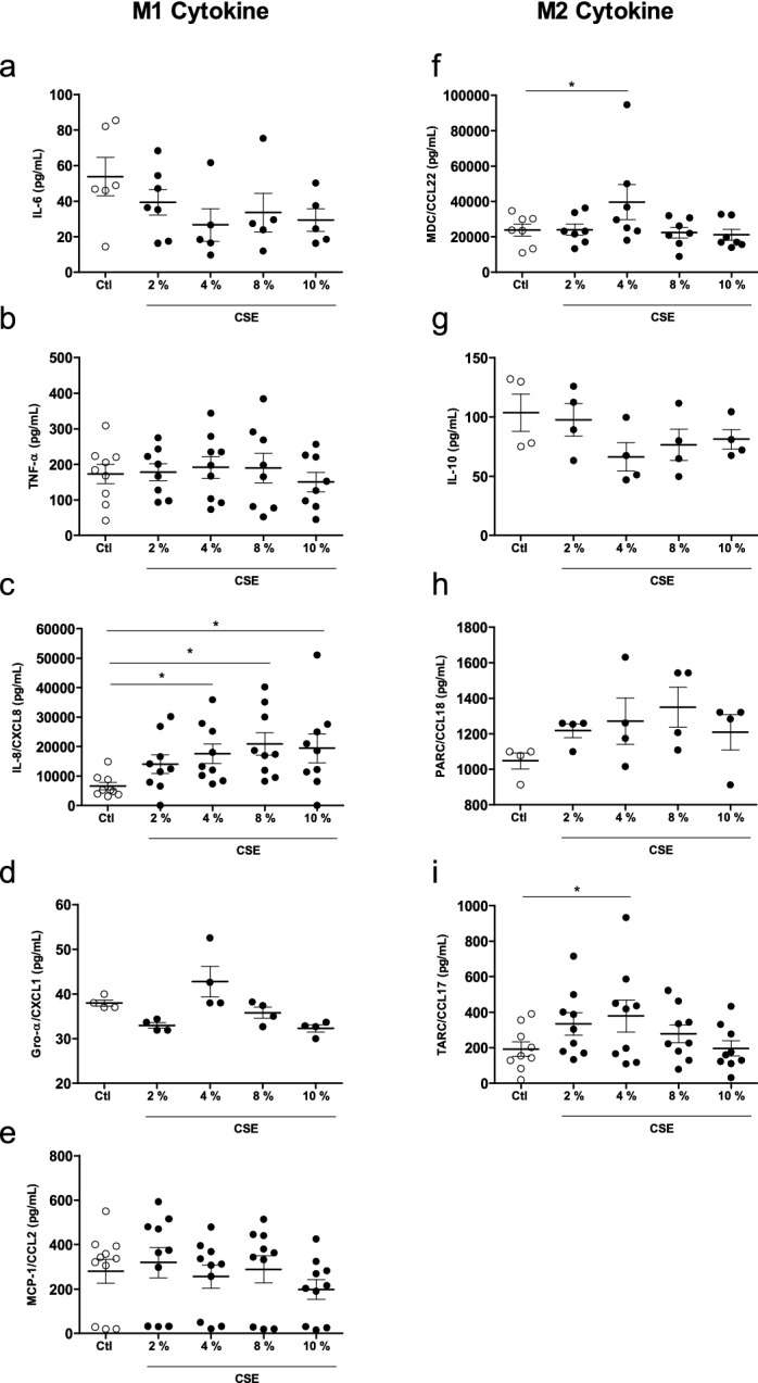
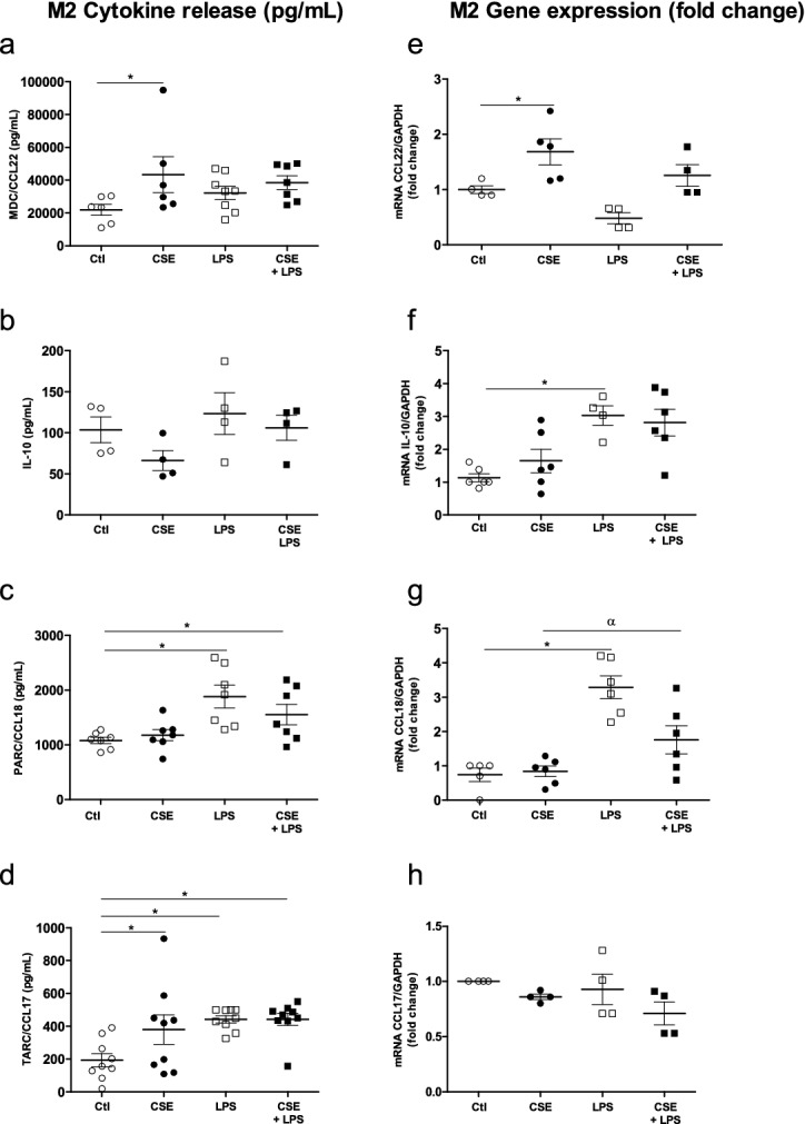
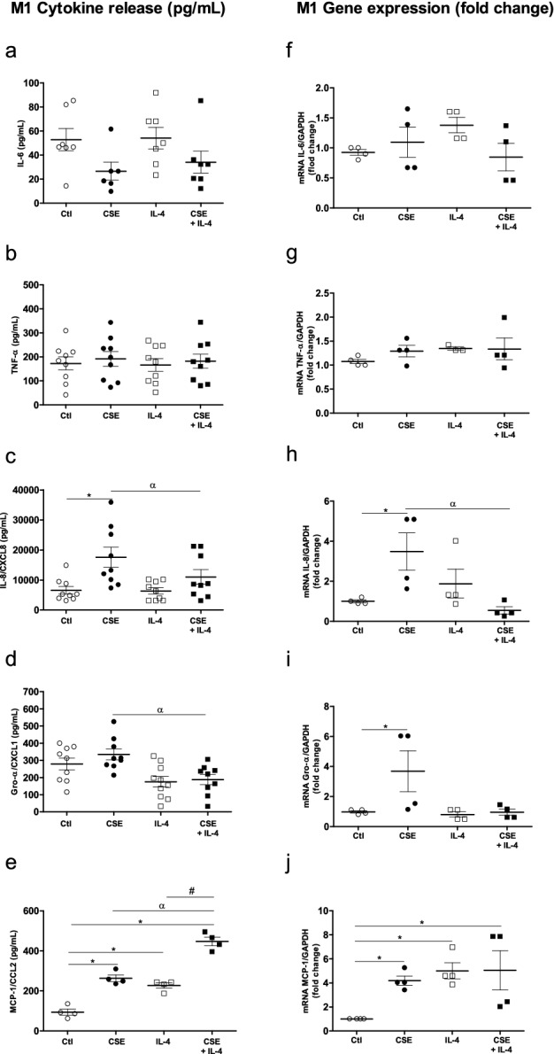
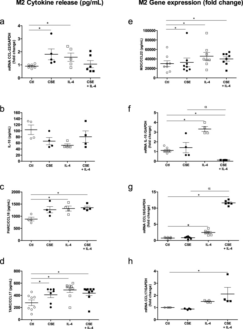
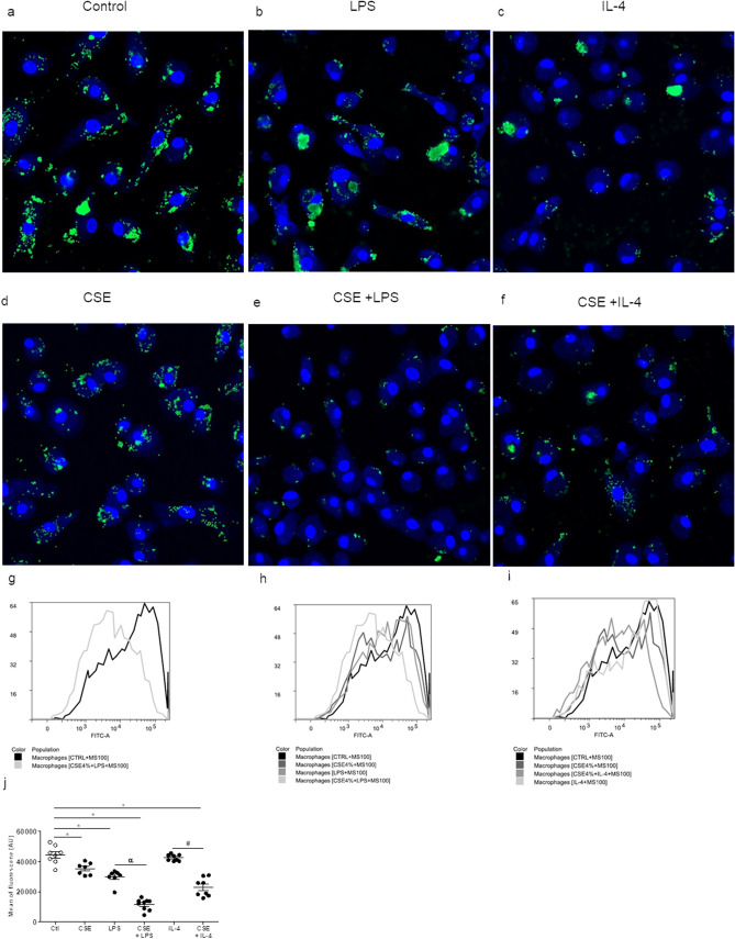
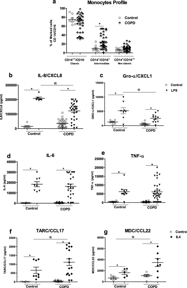
Cigarette smoke exposure (CS) is the main risk factor for chronic obstructive pulmonary disease (COPD). Macrophages have an important role in COPD because they release pro-inflammatory and anti-inflammatory cytokines. The present study's we investigate the functional changes in macrophages and monocytes exposed to cigarette smoke extract (CSE). Herein, using human monocyte-derived macrophages (MDMs) from healthy donors and we found that CSE was not associated with significant changes in the production of pro inflammatory cytokines by MDMs. In contrast, exposure to CSE suppressed the production of IL-6 and Gro-a/CXCL1 by LPS-stimulated-MDMs, but had an additive effect on the release of IL-8/CXCL8 and MCP1/CCL2. However, CSE exposure was associated with greater production, TARC/CCL-17 and CCL22/MDC. Moreover, MDMs displayed a lower uptake capacity after CSE exposure. We identify, for what is to our knowledge the first time that monocytes from patients with COPD produced less IL-8/CXCL8 and Gro-α/CXCL1 after LPS stimulation and produced higher levels of TARC/CCL17 and MDC/CCL-22 after IL-4 stimulation. Our present results highlighted a skewed immune response, with an imbalance in M1 vs. M2 cytokine production. In conclusion, exposure to CS has contrasting, multifaceted effects on macrophages and monocytes. Our data may provide a better understanding of the mechanisms underlying COPD.


Conflict of interest statement

The authors declare no competing interests

Figures

Figure 1
Figure 1
Effects of CSE on cytokine production by MDMs. The MDMs were incubated with medium alone (control) or with different concentration of CSE (2–10%) for 24 h. The culture supernatants were collected, and the concentrations of IL-6, TNF-α, IL-8/CXCL8, Gro-α/CXCL-1, MCP-1/CCL-2, MDC/CCL22, IL-10, PARC/CCL8, and TARC/CCL17 were measured using an ELISA. The data were assessed in an ANOVA, followed by a Bonferroni post-test. The data correspond to the mean ± SEM of 4–10 donors. *p < 0.05, compared with the control.
Figure 2
Figure 2
Effects of a combination of CSE and LPS on M1 cytokine production and expression by MDMs. The MDMs were incubated with medium alone (control), 4% CSE, 0.1 µg/mL LPS or 4% CSE + 0.1 µg/mL LPS for 2 h or 24 h. The culture supernatants were collected, and the concentrations of TNF-α, IL6, IL-8/CXCL8, Gro-α/CXCL1 and MCP-1/CCL-2 were measured using an ELISA (ae). MDMs were stimulated for 2 h, and mRNA expression was determined using RT-PCR (fj). The results were normalized against expression of the GAPDH gene. The data were assessed in an ANOVA, followed by a Bonferroni post-test. The data correspond to the mean ± SEM of 4–9 donors. *p < 0.05 compared with the control; αp < 0.05 compared with 4% CSE; #p < 0.05, compared with 0.1 µg/mL LPS.
Figure 3
Figure 3
Effects of a combination of CSE and LPS on M2 cytokine production and expression by MDMs. The MDMs were incubated with medium alone (control), 4% CSE, 0.1 µg/mL LPS, or 4% CSE + LPS for 24 h. The culture supernatants were collected, and the concentrations of MDC/CCL22, IL-10, PARC/CCL8 and TARC/CCL17 were measured using an ELISA (ad). MDMs were stimulated for 2 h, and mRNA expression was then determined using RT-PCR (eh). The results were normalized against expression of the GAPDH gene. The data were assessed in an ANOVA, followed by a Bonferroni post-test. The data correspond to the mean ± SEM of 4 to 9 donors. *p < 0.05, compared with the control; αp < 0.05, compared with 4% CSE; #p < 0.05, compared with 0.1 µg/mL LPS.
Figure 4
Figure 4
Effects of a combination of CSE and IL-4 on M1 cytokine production and expression by MDMs. The MDMs were incubated with medium alone (control), 4% CSE, 10 ng/mL IL-4 or CSE + IL-4 for 24 h. The culture supernatants were collected, and the concentrations of IL-6, TNF-α, IL-8/CXCL8, Gro-α/CXCL1 and MCP-1/CCL-2 were measured using an ELISA (ae). mRNA expression was then determined using RT-PCR (fj). The results were normalized against expression of the GAPDH gene. The data were assessed in an ANOVA, followed by a Bonferroni post-test. The data correspond to the mean ± SEM of 9 donors. *p < 0.05 compared with the control; αp < 0.05, compared with 4% CSE; #p < 0.05, compared with IL-4.
Figure 5
Figure 5
Effects of a combination of CSE and IL-4 on M2 cytokine production and expression by MDMs. The MDMs were incubated with medium alone (control), 4% CSE, 10 ng/mL IL-4 or 4% CSE + IL-4 10 µg/mL for 24 h. The culture supernatants were collected and the concentrations of MDC/CCL22, TARC/CCL17, IL-10, and PARC/CCL8 were measured using an ELISA (ad). mRNA expression was then determined using RT-PCR (ei). The results were normalized against expression of the GAPDH gene. The data were assessed in an ANOVA, followed by a Bonferroni post-test. The data correspond to the mean ± SEM of 9 donors. *p < 0.05 compared with the control; αp < 0.05, compared with 4% CSE; #p < 0.05 compared with 10 ng/mL IL-4.
Figure 6
Figure 6
Effects of CSE, CSE + LPS and CSE + IL-4 on microsphere uptake by MDMs. After 24 h of exposure to CSE, the culture medium was renewed with medium containing fluorescent microspheres (size: 100 nm) and incubated overnight. After incubation, the culture medium was discarded, and the cell monolayers was fixed with paraformaldehyde prior to observation under the confocal microscope (af). The fluorescence emitted by microspheres (on channel FL1-H) inside cells was analyzed using CellQuest cytometry software (gi). The intrinsic FL1-H fluorescence of MDMs was measured in the absence of microspheres. The data correspond to the mean ± SEM of representative experiment of one the 4 donors. *p < 0.05, compared with the control; αp < 0.05, compared with LPS; #p < 0.05, compared with CSE + IL-4.
Figure 7
Figure 7
Subpopulations of peripheral blood monocytes in healthy subjects and patients with COPD and the cells’ ability to release cytokines after M1 or M2 activation. Subpopulations of monocytes were determined by flow cytometry. The monocytes were stained for anti-CD14, anti-CD16 and anti-HLADR. The data are presented as the percentages of total classical (CD14++/CD16), intermediate (CD14+/CD16+) and non-classical (CD14+/CD16++) monocytes for each group (a). The total monocyte fraction was incubated with medium alone (control), 0.1 µg/mL LPS or 10 ng/mL IL-4 for 24 h. The culture supernatants were collected, and the concentrations of IL-8/CXCL8, IL-6, Gro-α/CXCL1, TNF-α, MDC/CCL22, and TARC/CCL17 and were measured using an ELISA (bg). The data were assessed in an ANOVA, followed by a Bonferroni post-test. The data correspond to the mean ± SEM of 8 to 37 donors. *p < 0.05, compared with the control; αp < 0.05, compared with LPS or IL-4.

Similar articles

Cited by

References

    1. Vogelmeier CF, et al. Global strategy for the diagnosis, management, and prevention of chronic obstructive lung disease 2017 report: GOLD executive summary. Arch. Bronconeumol. 2017;53:128–149. - PubMed
    1. Global initiative for chronic Obstructive Lung Disease. Global strategy for the diagnosis, management, and prevention of chronic obstructive pulmonary disease. https://goldcopd.org/wp-content/uploads/2018/11/GOLD-2019-v1.7-FINAL-14N.... - PubMed
    1. Buhl R, et al. Guideline for the diagnosis and treatment of asthma—Guideline of the German Respiratory Society and the German Atemwegsliga in cooperation with the Paediatric Respiratory Society and the Austrian Society of Pneumology. Pneumologie. 2017;71:e3. doi: 10.1055/a-0790-0021. - DOI - PubMed
    1. Barnes PJ. Inflammatory mechanisms in patients with chronic obstructive pulmonary disease. J. Allergy Clin. Immunol. 2016;138:16–27. doi: 10.1016/j.jaci.2016.05.011. - DOI - PubMed
    1. Herr C, et al. Suppression of pulmonary innate host defence in smokers. Thorax. 2009;64:144–149. doi: 10.1136/thx.2008.102681. - DOI - PubMed

Publication types

MeSH terms