

In vitro selection for adhesion of Plasmodium falciparum-infected erythrocytes to ABO antigens does not affect PfEMP1 and RIFIN expression
- PMID: 32732983
- PMCID: PMC7393120
- DOI: 10.1038/s41598-020-69666-9
In vitro selection for adhesion of Plasmodium falciparum-infected erythrocytes to ABO antigens does not affect PfEMP1 and RIFIN expression
Abstract
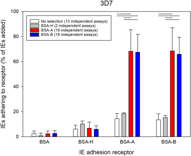
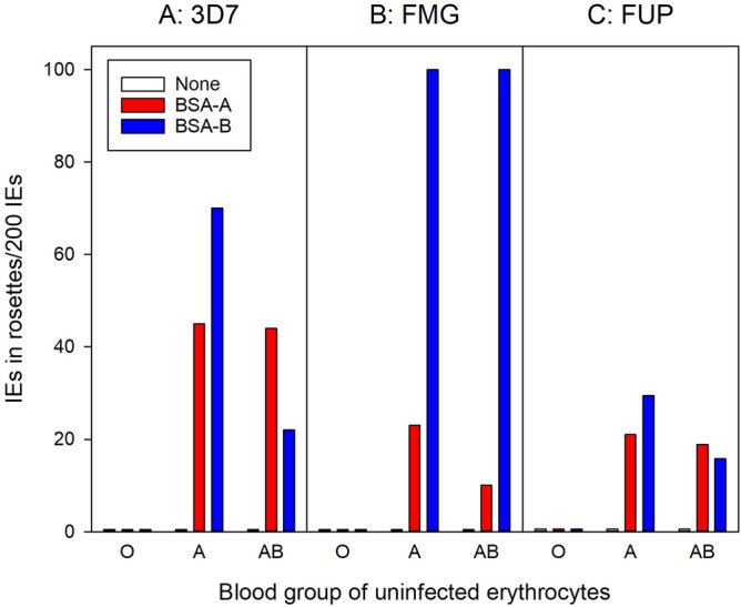
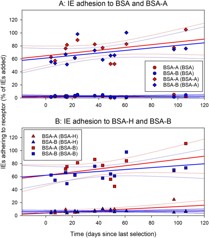
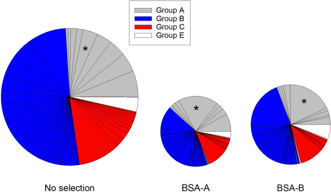
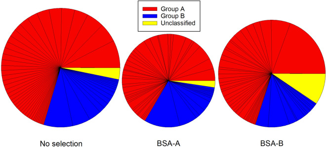
Plasmodium falciparum causes the most severe form of malaria in humans. The adhesion of the infected erythrocytes (IEs) to endothelial receptors (sequestration) and to uninfected erythrocytes (rosetting) are considered major elements in the pathogenesis of the disease. Both sequestration and rosetting appear to involve particular members of several IE variant surface antigens (VSAs) as ligands, interacting with multiple vascular host receptors, including the ABO blood group antigens. In this study, we subjected genetically distinct P. falciparum parasites to in vitro selection for increased IE adhesion to ABO antigens in the absence of potentially confounding receptors. The selection resulted in IEs that adhered stronger to pure ABO antigens, to erythrocytes, and to various human cell lines than their unselected counterparts. However, selection did not result in marked qualitative changes in transcript levels of the genes encoding the best-described VSA families, PfEMP1 and RIFIN. Rather, overall transcription of both gene families tended to decline following selection. Furthermore, selection-induced increases in the adhesion to ABO occurred in the absence of marked changes in immune IgG recognition of IE surface antigens, generally assumed to target mainly VSAs. Our study sheds new light on our understanding of the processes and molecules involved in IE sequestration and rosetting.
Conflict of interest statement
The authors declare no competing interests.
Figures
Similar articles
-
α2-Macroglobulin Can Crosslink Multiple Plasmodium falciparum Erythrocyte Membrane Protein 1 (PfEMP1) Molecules and May Facilitate Adhesion of Parasitized Erythrocytes.PLoS Pathog. 2015 Jul 2;11(7):e1005022. doi: 10.1371/journal.ppat.1005022. eCollection 2015 Jul. PLoS Pathog. 2015. PMID: 26134405 Free PMC article.
-
Rifins, rosetting, and red blood cells.Trends Parasitol. 2015 Jul;31(7):285-6. doi: 10.1016/j.pt.2015.04.009. Epub 2015 May 7. Trends Parasitol. 2015. PMID: 25959958
-
Surface antigens of Plasmodium falciparum-infected erythrocytes as immune targets and malaria vaccine candidates.Cell Mol Life Sci. 2014 Oct;71(19):3633-57. doi: 10.1007/s00018-014-1614-3. Epub 2014 Apr 2. Cell Mol Life Sci. 2014. PMID: 24691798 Free PMC article. Review.
-
Three Is a Crowd - New Insights into Rosetting in Plasmodium falciparum.Trends Parasitol. 2017 Apr;33(4):309-320. doi: 10.1016/j.pt.2016.12.012. Epub 2017 Jan 18. Trends Parasitol. 2017. PMID: 28109696 Review.
-
Multiple adhesive phenotypes linked to rosetting binding of erythrocytes in Plasmodium falciparum malaria.Infect Immun. 1998 Jun;66(6):2969-75. doi: 10.1128/IAI.66.6.2969-2975.1998. Infect Immun. 1998. PMID: 9596774 Free PMC article.
Cited by
-
PfEMP1 and var genes - Still of key importance in Plasmodium falciparum malaria pathogenesis and immunity.Adv Parasitol. 2024;125:53-103. doi: 10.1016/bs.apar.2024.02.001. Epub 2024 Mar 23. Adv Parasitol. 2024. PMID: 39095112 Free PMC article. Review.
References
-
- World Health Organization, N. World malaria report 2018 (2018).
-
- David PH, Handunnetti SM, Leech JH, Gamage P, Mendis KN. Rosetting: a new cytoadherence property of malaria-infected erythrocytes. Am. J. Trop. Med. Hyg. 1988;38:289–297. - PubMed
-
- Handunnetti SM, David PH, Perera KLRL, Mendis KN. Uninfected erythrocytes form 'rosettes' around Plasmodium falciparum infected erythrocytes. Am. J. Trop. Med. Hyg. 1989;40:115–118. - PubMed
-
- Udomsangpetch R, Thanikkul K, Pukrittayakamee S, White NJ. Rosette formation by Plasmodium vivax. Trans. R. Soc. Trop. Med. Hyg. 1995;89:635–637. - PubMed
Publication types
MeSH terms
Substances
LinkOut - more resources
Full Text Sources
