. 2020 Jul 30;10(1):12803.
doi: 10.1038/s41598-020-69638-z.

Simultaneous time-varying viscosity, elasticity, and mass measurements of single adherent cancer cells across cell cycle

Affiliations

Simultaneous time-varying viscosity, elasticity, and mass measurements of single adherent cancer cells across cell cycle

Olaoluwa O Adeniba et al. Sci Rep. .

Abstract

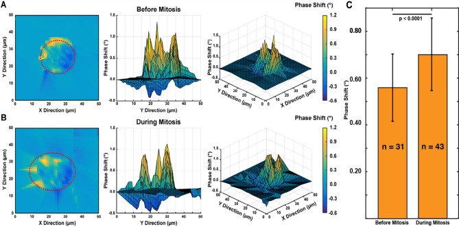
Biophysical studies on single cells have linked cell mechanics to physiology, functionality and disease. Evaluation of mass and viscoelasticity versus cell cycle can provide further insights into cell cycle progression and the uncontrolled proliferation of cancer. Using our pedestal microelectromechanical systems resonant sensors, we have developed a non-contact interferometric measurement technique that simultaneously tracks the dynamic changes in the viscoelastic moduli and mass of adherent colon (HT-29) and breast cancer (MCF-7) cells from the interphase through mitosis and then to the cytokinesis stages of their growth cycle. We show that by combining three optomechanical parameters in an optical path length equation and a two-degree-of-freedom model, we can simultaneously extract the viscoelasticity and mass as a function of the nano-scaled membrane fluctuation of each adherent cell. Our measurements are able to discern between soft and stiff cells across the cell cycle and demonstrated sharp viscoelastic changes due to cortical stiffening around mitosis. Cell rounding before division can be detected by measurement of mechanical coupling between the cells and the sensors. Our measurement device and method can provide for new insights into the mechanics of single adherent cells versus time.


Conflict of interest statement

The authors declare no competing interests.

Figures

Figure 1
Figure 1
Overview of measurement scheme. (A) Summary of the vibration induced phase shift (Δϕ),amplitude ratio, ΔA, and frequency shift (Δf) relate to the viscoelastic properties and mass of the cell. These parameters are extracted during the vibration of a cell-loaded sensor while the cell cycle stage progression is being observed through phases G1–S–G2–M. (B) Cross-sectional figures elucidating the vibration induced phase shift, Δϕ of a targeting signal beam, ϕ1 and ϕ2 on an empty sensor; inside and outside a rigid cell; and inside and outside of a viscoelastic cell; compared to a reference beam, ϕ3. G*(є) represents the cell dynamic moduli (viscoelastic moduli) as a function of lateral position, є, which tracks the cell heights oscillation. (C) Model of sensor-cell system as a 2-DOF suspended mass model where the cell mass (m2) is considered a Kelvin-Voigt viscoelastic solid with elastic stiffness (k2) and viscous coefficient (c2) connected to the sensor, and the sensor mass (m1) is connected to the fixed substrate by a second Kelvin-Voigt spring-damper (k1, c1). The model assumes an oscillatory force F(t) applied to the sensor mass. (D) Cell physiological transition from initial adhesion through cell rounding/division to reattachment to a patterned surface.
Figure 2
Figure 2
Mechanical viscoelastic properties change as cells grow. (A) Experimental timeline highlighting the transition of cell optomechanical measurement on sensors with respect to the LDV position while carrying out fluorescence imaging. (BC) Cell mass, elasticity, and viscosity versus time for both (B) HT-29 and (C) MCF-7. For single cell growth analysis of HT-29 and MCF-7 cells, the mass and stiffness data was analyzed for the interphase (i, ii), prior to mitosis (iii) and after a mitotic event (iv and v). Division is shown with the individual cells and daughter cells. These parameters are extracted during the vibration of a cell-loaded sensor while the cell cycle stage progression is being observed through phases G1 (Red)–S (Orange)–G2 (Green)–M (Green) using FUCCI cell cycle reporter. (DE) Comparison of average interphase elasticity and viscosity values for both (D) HT-29 (n = 9) and (E) MCF-7 (n = 5) cells during interphase (average) versus pre-mitosis (iii). Data presented as a mean ± standard deviation.
Figure 3
Figure 3
RhoA significantly increases viscoelasticity. (A) Time-Varying Mass, Elasticity and Viscosity measurement of RhoActivator—mediated HT-29 cells (treated) and unmodified HT-29 cells (untreated) measured by our MEMS resonator. Plot show a significant increase in cell viscoelastic moduli of modified HT-29 cells due to phosphorylation of actin. (B) Comparison between plot showing a significant increase in cell viscoelastic moduli of modified MCF-7 cells due to phosphorylation of actin fibres. Dashed lines are used to trace the mass and viscoelastic values per cell cycle stage. MCF-7 remains in the G2 checkpoint phase before apoptosis. (CD) Comparison of average Elasticity and Viscosity values for RhoActivator—mediated (treated) and untreated (D) HT-29 (treated: n = 4, untreated: n = 9) and (E) MCF-7 (treated: n = 4, untreated: n = 5) cells during interphase. Plot shows a significant difference in viscoelastic moduli for treated cells versus untreated cells. Data presented as a mean ± standard deviation.
Figure 4
Figure 4
HT-29 (colon) and MCF-7 (breast) cancer cell spreading response to Rho Activator. Quantification for HT-29 and MCF-7 treated and untreated cells with the Rho Activator for their: (A) Cytoskeleton Spreading (B) Nuclear Spreading. Plot shows cell area (spreading) of both cell lines increased with the addition of the Rho activator, which is indicative of increased actin polymerization. Data presented as a mean ± standard deviation. (C) Representative images of HT-29 and MCF-7 treated and untreated with the Rho Activator comparing cytoskeletal and nuclear spread.
Figure 5
Figure 5
Scanning measurement maps of vibration induced phase shifts (VIPS) of an HT-29 cell. These maps indicate stiffness differences of a HT-29 cell at different stages of the cell cycle. Top, side, and 3D views of the same live cell both (A) prior to and (B) during mitosis. Prior to mitosis, we observed an increase in stiffness and an average inside cell (dotted red lines) lower phase shift (VIPS) of 0.56 ± 0.21°. During mitosis, cells are partially detached—cell height oscillation increases (with softness); hence a higher inside cell (dotted red lines) VIPS of 0.75 ± 0.31° is observed. (C) Bar chart showing statistically significant differences (p < 0.0001) in the pre-mitosis (n = 31) and during-mitosis (n = 43) phase shift (VIPS) values. Data presented as a mean ± standard deviation.

References

    1. Corbin EA, Kong F, Lim T, King PW, Bashir R. Biophysical properties of human breast cancer cells measured using silicon MEMS resonators and atomic force microscopy. Lab Chip. 2014;15(3):839–47. - PubMed
    1. Cross SE, Jin Y-C, Rao J, Gimzewski JK. Nanomechanical analysis of cells from cancer patients. Nat. Nanotechnol. 2007;2:780–783. - PubMed
    1. Byun S, et al. Characterizing deformability and surface friction of cancer cells. Proc. Natl. Acad. Sci. 2013;110:7580–7585. - PMC - PubMed
    1. Xu W, Mezencev R, Kim B, Wang L, McDonald J, Sulchek T. Cell stiffness is a biomarker of the metastatic potential of ovarian cancer cells. PLoS ONE. 2012;7:e46609. - PMC - PubMed
    1. Cavallaro U, Christofori G. Cell adhesion and signalling by cadherins and Ig-CAMs in cancer. Nat. Rev. Cancer. 2004;4:118. - PubMed

Publication types