

Taxonomy and phylogeny of Sidera (Hymenochaetales, Basidiomycota): four new species and keys to species of the genus
- PMID: 32733146
- PMCID: PMC7360635
- DOI: 10.3897/mycokeys.68.53561
Taxonomy and phylogeny of Sidera (Hymenochaetales, Basidiomycota): four new species and keys to species of the genus
Abstract
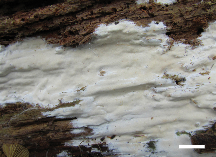
Sidera is a polypore genus with white to cream or buff basidiomata, whose species in Hymenochaetales are poorly known. We study the phylogeny and diversity of Sidera based on our recent collections from tropic and subtropic Asian-Pacific regions. Phylogenetic analyses based on the internal transcribed spacer (ITS) and nuclear large subunit (nLSU) ribosomal RNA gene regions indicate that ten terminal lineages are well supported within Sidera. Based on morphological examination and phylogeny, four new species, viz. Sidera minutissima, S. parallela, S. srilankensis and S. tenuis are described, and a new combination, Sidera minutipora, is proposed. All these species are illustrated. Sidera minutissima is characterized by tiny basidiomata with bluish pores when fresh, generative hyphae dominating at the dissepiment edges, the presence of cystidioles, and allantoid basidiospores measuring 3.8-4.4 × 0.9-1.3 μm. Sidera parallela differs from other poroid species in the genus by having parallel tramal hyphae in combination with lunate basidiospores measuring 2.8-3.3 × 0.9-1.2 μm. Sidera srilankensis have generative and skeletal hyphae co-dominating at the dissepiment edges, and lunate basidiospores measuring 3.5-4 × 1-1.3 μm. Sidera tenuis is distinguished by small pores (8-10 per mm) and relatively long allantoid basidiospores measuring 4.2-5 × 0.8-1 μm. Sidera minutipora is characterized by buff to olivaceous buff basidiomata when dry, 5-7 pores per mm, rosette-like crystals rare, and allantoid basidiospores measuring 3.7-4.3 × 1-1.3 μm. An identification key to all accepted species is provided.
Keywords: Rickenellaceae; Phylogeny; taxonomy; wood-rotting fungi.
Rui Du, Fang Wu, Genevieve M. Gates, Yu-Cheng Dai, Xue-Mei Tian.
Figures


References
-
- Anonymous (1969) Flora of British Fungi. Colour Identification Chart. Her Majesty´s Stationery Office, London.
-
- Bourdot H, Galzin A. (1928) Hyménomycètes de France. France, 764 pp.
-
- Buchanan PK, Ryvarden L. (1993) Type studies in the Polyporaceae 24. Species described by Cleland, Rodway and Cheel. Australian Systematic Botany 6: 215–235. 10.1071/SB9930215 - DOI
-
- Cunningham GH. (1965) Polyporaceae of New Zealand. Bulletin of the New Zealand Department Scientific and Industrial Research 64: 1–304.
-
- Cui BK, Li HJ, Ji X, Zhou JL, Song J, Si J, Yang ZL, Dai YC. (2019) Species diversity, taxonomy and phylogeny of Polyporaceae (Basidiomycota) in China. Fungal Diversity 97: 137–392. 10.1007/s13225-019-00427-4 - DOI
LinkOut - more resources
Full Text Sources