Review
. 2020 Jul 9:11:1395.
doi: 10.3389/fimmu.2020.01395. eCollection 2020.

Innate Immune Defense Mechanisms by Myeloid Cells That Hamper Cancer Immunotherapy

Affiliations
Review

Innate Immune Defense Mechanisms by Myeloid Cells That Hamper Cancer Immunotherapy

Els Lebegge et al. Front Immunol. .

Abstract

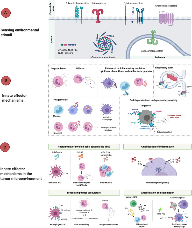
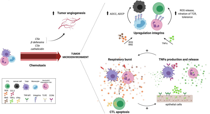
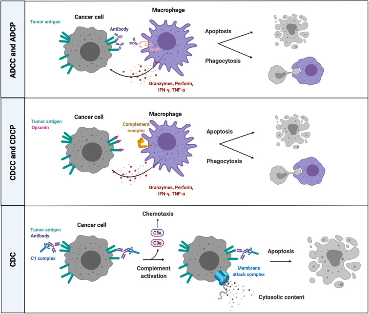
Over the past decade, cancer immunotherapy has been steering immune responses toward cancer cell eradication. However, these immunotherapeutic approaches are hampered by the tumor-promoting nature of myeloid cells, including monocytes, macrophages, and neutrophils. Despite the arsenal of defense strategies against foreign invaders, myeloid cells succumb to the instructions of an established tumor. Interestingly, the most primordial defense responses employed by myeloid cells against pathogens, such as complement activation, antibody-dependent cell cytotoxicity and phagocytosis, actually seem to favor cancer progression. In this review, we discuss how rudimentary defense mechanisms deployed by myeloid cells can promote tumor progression.

Keywords: cancer immunotherapy; immune suppression; immunotherapy resistance; innate immune response; tumor microenvironment; tumor-associated myeloid cells.


Figures

Figure 1
Figure 1
Linear representation of classical innate immunity in response to threats and in the TME. (A) PAMPs and DAMPs are recognized by surface-expressed, endosomal and cytosolic pattern recognition receptors (TLR, CLR, cytokine, chemokine receptors, NLRP3) which results in phenotypical changes that counteract ongoing threats or tissue damage. (B) Effector mechanisms that take place during inflammation are degranulation, NETosis, release of proinflammatory mediators, respiratory burst, phagocytosis and cell-dependent and -independent cytotoxicity. The net result is the recruitment of immunocompetent cells that mount an inflammatory reaction and potentially resolve the infection. (C) However, in the tumor microenvironment innate myeloid cells promote tumor progression through active recruitment to the TME in response to ß-defensins, cathelicidin, G-CSF, complement factors and chemokines. Once arrived in the TME, myeloid cells are activated and release proinflammatory mediators, which empowers tumor-associated inflammation. Activation of myeloid cells also allows for remodeling of the tissue vasculature and extracellular matrix, which also allows for cancer-cell invasion and metastasis. Furthermore, myeloid cells contribute to immunosuppression once activated by for example, upregulation of PD-L1 and IDO release during antibody-dependent phagocytosis of target cells or stimulatory cytokines (IFNγ). DC, dendritic cell; ECM, extracellular matrix; VEGFR2, vascular endothelial growth factor receptor 2; IFNγ, interferon gamma; ROS, reactive oxygen species; MDSC, myeloid-derived suppressor cell; ADCP, antibody-dependent cell-mediated phagocytosis; IDO, indoleamine 2,3-dioxygenase; COX2, cyclooxygenase 2; PGE2, prostaglandin E2; TNFα, tumor necrosis factor alpha; G-CSF, granulocyte colony stimulating factor; NOD, nucleotide-binding oligomerization domain; RIG, retinoic acid-inducible gene; NLRP, nucleotide-binding oligomerization domain; leucine-rich repeat and pyrin domain containing.
Figure 2
Figure 2
Cross talk between reoccurring innate effector mechanisms in the TME. Tumor-derived chemokines that are produced as a result of innate effector mechanisms including C5a, C3a, cathelicidin and ß-defensin, recruit myeloid cells to the TME. Tissue vasculature during chronic inflammation is maintained by complement anaphylatoxin C5a and beta-defensins. Anaphylatoxin C5a also recruits MDSCs with increased ROS and NRS production in the TME. Many innate pathways converge at the production of ROS and NOS in the TME. For example, TLR2 signaling increases the antigen presenting capacity of TAMs, which activates CTLs resulting in IFNγ release and subsequent ROS and NO release by TAMs. Neutrophil-derived ROS induces CTL apoptosis, while hydrogen peroxide released by TAMs, induces the expression of TNFα and TNFαR1 in surrounding epithelial cells. A positive feedback loop seems to exist between the respiratory burst and TNFα release, creating a potential cross talk between TAMs, neutrophils and epithelial cells in the TME. Furthermore, both ROS and TNFα also increases the expression of integrins, which increases cell-cell contact and facilitates cell-mediated killing via ADCC and ADCP either by performed by monocytes to kill cancer cells, or by MDSCs to suppress CTLs. ADCC, antibody-dependent cell-mediated cytotoxicity; MDSC, myeloid-derived suppressor cell; ADCP, antibody-dependent cell-mediated phagocytosis; ROS, reactive oxygen species; RNS, reactive nitrogen species; TNFα, tumor necrosis factor alpha; TNFαR1, tumor necrosis factor alpha receptor 1; CTL, cytotoxic T lymphocyte; IFNγ, interferon gamma; TCR, T-cell receptor; TAM, tumor-associated macrophage; CTL, cytotoxic T lymphocyte; TME, tumor microenvironment; TLR2, Toll-like receptor 2; TAM, tumor-associated macrophage.
Figure 3
Figure 3
Cell-dependent and -independent effector mechanisms of complement activation and FcR-mediated killing. Complement factor- and antibody-opsonized cancer cells can be eliminated through cell-dependent and cell-independent effector mechanisms. CRs and FcRs on phagocytes bind opsonins and antibodies, respectively, on the surface of targeted cancer cells, followed by phagocytosis and/or release of lytic enzymes (granzyme B, perforins) and proinflammatory mediators (TNFα, IFNγ). The classical pathway of complement activation mediates a cell-independent form of lytic cell death by introducing a MAC in the membrane of antibody opsonized target cells that are recognized by complement C1 complex. ADCC, antibody-dependent cell-mediated cytotoxicity; ADCP, antibody-dependent cell-mediated phagocytosis; CDC, complement-dependent cytotoxicity; CDCC, complement-dependent cell-mediated cytotoxicity; CDCP, complement-dependent cell-mediated phagocytosis; CRs, complement receptors; IFNγ, interferon gamma; TNFα, tumor necrosis factor alpha; C5a, complement factor 5a; C3a, complement factor C3a; FcγR, crystallizable fragment receptor gamma; C1, complement factor.

References

    1. Chen DS, Mellman I. Oncology meets immunology: the cancer-immunity cycle. Immunity. (2013) 39:1–10. 10.1016/j.immuni.2013.07.012 - DOI - PubMed
    1. Woo S-R, Corrales L, Gajewski TF. Innate immune recognition of cancer. Annu Rev Immunol. (2015) 33:445–74. 10.1146/annurev-immunol-032414-112043 - DOI - PubMed
    1. Colotta F, Allavena P, Sica A, Garlanda C, Mantovani A. Cancer-related inflammation, the seventh hallmark of cancer: links to genetic instability. Carcinogenesis. (2009) 30:1073–81. 10.1093/carcin/bgp127 - DOI - PubMed
    1. Balkwill F, Charles KA, Mantovani A. Smoldering and polarized inflammation in the initiation and promotion of malignant disease. Cancer Cell. (2005) 7:211–17. 10.1016/j.ccr.2005.02.013 - DOI - PubMed
    1. De Visser KE, Eichten A, Coussens LM. Paradoxical roles of the immune system during cancer development. Nat Rev Cancer. (2006) 6:24–37. 10.1038/nrc1782 - DOI - PubMed

Publication types