. 2020 Jul 8:11:1430.
doi: 10.3389/fimmu.2020.01430. eCollection 2020.

Characterization of a Novel Compound That Stimulates STING-Mediated Innate Immune Activity in an Allele-Specific Manner

Affiliations

Characterization of a Novel Compound That Stimulates STING-Mediated Innate Immune Activity in an Allele-Specific Manner

Jinu Abraham et al. Front Immunol. .

Abstract

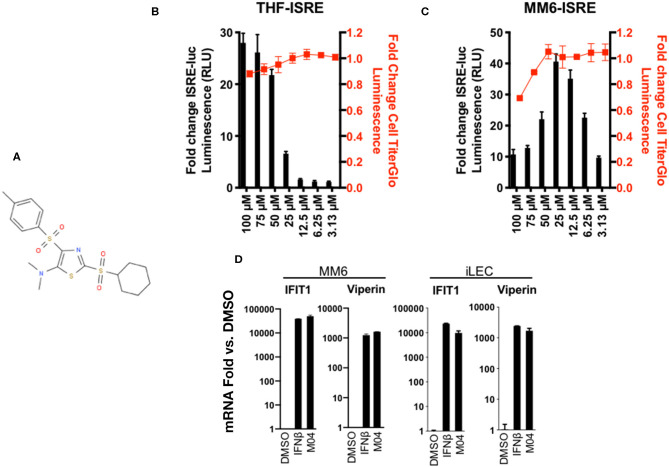
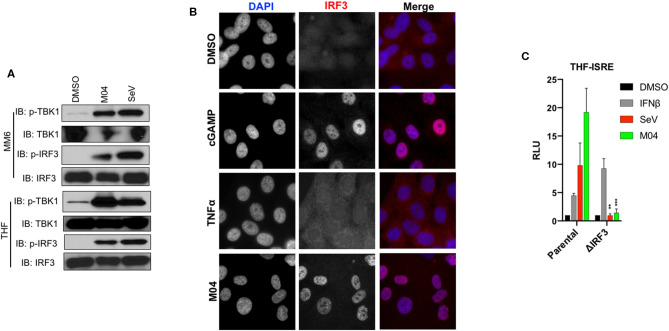
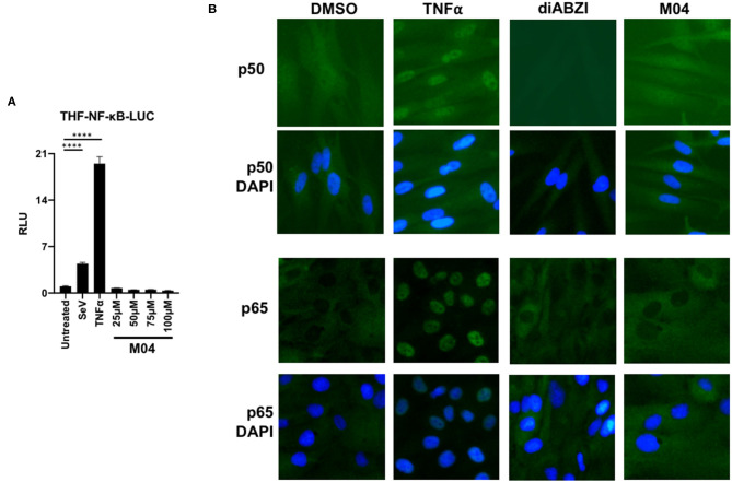
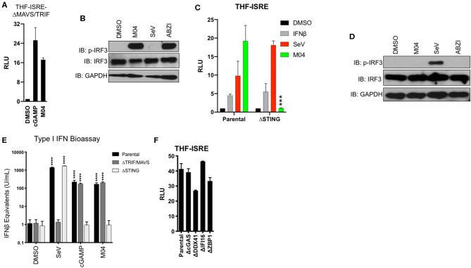
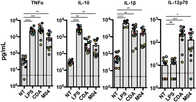
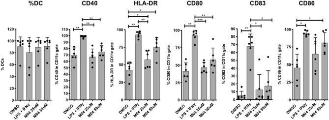
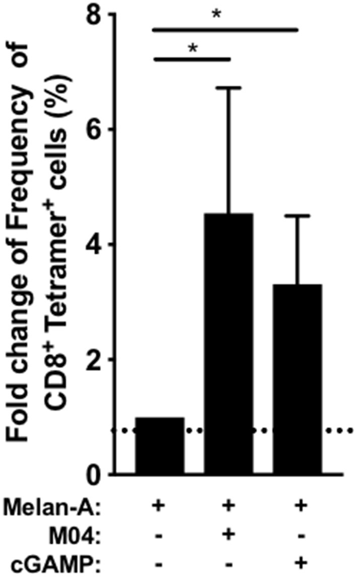
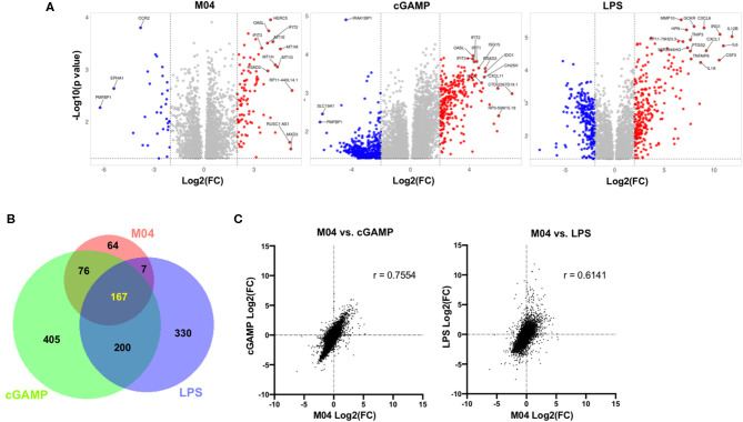
The innate immune response to cytosolic DNA involves transcriptional activation of type I interferons (IFN-I) and proinflammatory cytokines. This represents the culmination of intracellular signaling pathways that are initiated by pattern recognition receptors that engage DNA and require the adaptor protein Stimulator of Interferon Genes (STING). These responses lead to the generation of cellular and tissue states that impair microbial replication and facilitate the establishment of long-lived, antigen-specific adaptive immunity. Ultimately this can lead to immune-mediated protection from infection but also to the cytotoxic T cell-mediated clearance of tumor cells. Intriguingly, pharmacologic activation of STING-dependent phenotypes is known to enhance both vaccine-associated immunogenicity and immune-based anti-tumor therapies. Unfortunately, the STING protein exists as multiple variant forms in the human population that exhibit differences in their reactivity to chemical stimuli and in the intensity of molecular signaling they induce. In light of this, STING-targeting drug discovery efforts require an accounting of protein variant-specific activity. Herein we describe a small molecule termed M04 that behaves as a novel agonist of human STING. Importantly, we find that the molecule exhibits a differential ability to activate STING based on the allelic variant examined. Furthermore, while M04 is inactive in mice, expression of human STING in mouse cells rescues reactivity to the compound. Using primary human cells in ex vivo assays we were also able to show that M04 is capable of simulating innate responses important for adaptive immune activation such as cytokine secretion, dendritic cell maturation, and T cell cross-priming. Collectively, this work demonstrates the conceivable utility of a novel agonist of human STING both as a research tool for exploring STING biology and as an immune potentiating molecule.

Keywords: STING; adjuvant; cytokine; cytosolic DNA sensing; interferon.


Figures

Figure 1
Figure 1
Dose-Dependent Activation of Type I Interferon-Mediated Signaling and Cytotoxicity of M04 in Human Cells. (A) Chemical structure of 2-(cyclohexylsulfonyi)-N,N-dimethyl-4-tosylthiazol-5-amine (”M04"); (B) ISRE-dependent expression of Luciferase (LUC) as well as relative cellular viability as determined by Cell Titer Glo in THF (B) and MM6 (C) cells exposed to M04 at indicated concentrations (μM) for 8 h (RLU) or 24 h (Cell Titer Glow). Values presented are mean fold change ± SD relative to cells treated with 1% DMSO (RLU; black bars; left y-axis). Cell viability data are expressed as relative signal detected in DMSO-treated cells (red squares; right axis). Values displayed are based on four replicates; (D) Fold changes of IFIT1 or Viperin mRNA relative to 1% DMSO treatment in immortalized lymphatic endothelial cells (iLEC) or MM6 following 8 h exposure to 1000U/mL IFNβ or 50 μM M04 as indicated. Presented values represent average ± SD mRNA fold changes relative to cells exposed to untreated cells from duplicate experiments.
Figure 2
Figure 2
M04 induces canonical activation of IRF3 which is essential to reporter signal generated by the compound. (A) Immunoblot showing phosphorylation status of TBK1 Ser172 and IRF3 Ser386 as well as corresponding total protein levels in MM6 cells (left) and THF (right) exposed for 4 h to 1% DMSO, 50 μM M04, or 1,000 HAU/mL SeV as indicated; (B) Indirect immunofluorescence showing subcellular localization of IRF3 in THF exposed for 4 h to 1% DMSO, transfected 2'3'cGAMP (10 μg/mL), 100 ng/mL TNFα, or 50 μM M04; (C) Reporter assay illustrating IFN-dependent LUC induction following overnight treatment with 1% DMSO, 1,000 U/mL IFNβ, 1,000 HAU/mL SeV, or 50 μM M04 in parental cells as well as those from which IRF3 was deleted as indicated. Data presented are mean ± SD relative luminescence units (RLU) using signal from DMSO-treated cells based on quadruplicate measurements. Student's T-test was used to compare RLU in the parental and ΔIRF3 cells **P < 0.01; ***P < 0.001.
Figure 3
Figure 3
M04 does not Activate NF-κB-Dependent Processes. (A) Reporter assay using THF cells responsive to activated NF-κB showing induction of LUC expression following 8 h treatment with 160 HAU/mL SeV, 10 ng/mL TNFα, or the indicated concentration of M04. Values displayed are average fold changes (±SD) based on four replicates compared to DMSO-treated cells; (B) Indirect immunofluorescence showing subcellular localization of NF-KB P65 subunit in THF exposed for 4 h to DMSO, 100 ng/mL TNFα, or 75 μM M04. Statistical significance between treated and untreated cells was then calculated using Student's T-test ****p < 0.0001.
Figure 4
Figure 4
Innate Activation by M04 requires STING but not MAVS, TRIF, or cytosolic DNA PRRs. (A) Reporter assay illustrating IFN-dependent LUC induction in THF-ISRE-ΔMAVS/TRIF following overnight treatment with 1% DMSO, transfected cGAMP (10 μg/mL), or 75 μM M04. Data presented are mean ± SD relative luminescence units (RLU) using signal from DMSO-treated cells as the basis (n = 4 treatments); (B) Immunoblot showing phosphorylation status of IRF3 Ser386, total IRF3, and GAPDH in THF-ISRE-ΔMAVS/TRIF following 8 h treatment with 1% DMSO, 75 μM M04, 1,000 HAU/mL SeV or 25 μM ABZI as indicated; (C) Reporter assay illustrating IFN-dependent LUC induction in THF-ISRE-ΔSTING following overnight treatment with 1% DMSO, 1,000 U/mL IFNβ, 1,000 HAU/mL SeV, or 75 μM M04. Data presented are mean RLU ± SD as described above; Student's T-test was used to compare RLU ***p < 0.001; (D) Immunoblot showing phosphorylation status of IRF3 Ser386, total IRF3 in THF-ISRE-ΔSTING following 4 h treatment with 1% DMSO, 50 μM M04, 1,000 HAU/mL SeV or 25 μM ABZI as indicated; (E) Secretion of bioactive type I IFN from parental THF as well as THF-ISRE-ΔMAVS/TRIF and THF-ISRE-ΔSTING treated in triplicate overnight with 1% DMSO, 1,000 HAU/mL SeV, transfected cGAMP (10 μg/mL), or 75 μM M04. Data are expressed as mean concentrations ± SD for IFNβ equivalent units. Statistical significance between treated and untreated cells of similar genetic background was calculated using Student's T-test. ****p < 0.0001; (F) Reporter assay from WT parental THF-ISRE cells as well as from cells from which indicated dsDNA-specific PRRs were deleted. Values presented are mean fold changes ± SD for duplicates relative to the value for DMSO-treated cells.
Figure 5
Figure 5
M04 Induces Canonical STING Activation. (A) lmmunoblot showing phosphorylation status of STING Ser366 as well as total STING in THF and MM6 following 4 h treatment with 1% DMSO, 50μM M04, 1,000 HAU/mL SeV or 25 μM ABZI as indicated; (B) Indirect immunofluorescence showing subcellular localization of Golgi marker GM130 and STING in THF exposed for 4 h to DMSO, transfected cGAMP (10 μg/mL), 100 ng/mL TNFα, or 50 μM M04; (C) Melting temperature shifts for human STING-CTD in the presence of DMSO, 75 μM M04, or 100 μM 2'3' cGAMP. Data presented are SYPRO orange relative fluorescent units (RFU).
Figure 6
Figure 6
Responsiveness to M04 can be Conferred through Introduction of WT STING Allelic Variant. (A) Reporter assay illustrating IFN-dependent LUC induction in THP-1-ISG-Lucia following overnight treatment with 1% DMSO, 1,000 HAU/mL SeV, 1,000 U/mL IFNβ, 75 μM TRIF agonist AV-C, 25 μM ABZI, or 75 μM M04. Data presented are mean ± SD relative luminescence units (RLU) using signal from DMSO-treated cells as the basis (n = 4 treatments). Student's T-test was used to compare RLU ***p < 0.001, ****p < 0.0001; (B) lmmunoblot showing phosphorylation status of IRF3 Ser386 and total IRF3 in THP-1 whole cell lysates following 4 h treatment with 1% DMSO, 50 μM M04, 1,000 HAU/mL SeV, 25 μM ABZI, or 10 μg/mL cGAMP as indicated; (C) lmmunoblot showing expression of endogenous or ectopically expressed WT hSTING in THP-1 as indicated. (D) Immunoblot showing phosphorylation status of IRF3 Ser386 in THP-1 cells from which endogenous STING was deleted and WT STING stably introduced following indicated treatment as described above.
Figure 7
Figure 7
Transient transfection of vectors encoding WT and R232H but not HAQ hSTING confer responsiveness to M04. (A) lmmunoblot from HEK293T whole cell lysates showing expression of indicated STING variants following transient transfection, S386 phosphorylation status of IRF3 and total IRF3. Cells were left untreated or exposed to 75 μM M04, 100 nM diABZI, or 10 μg/mL cGAMP as indicated; (B) Reporter assay using cells (n = 4) treated as described in (A). Values displayed are mean fold changes ± SD relative to cells transfected with empty vector; (C) Expression of IFIT1 and Viperin mRNA as determined by qPCR in parental A549 cells as well as those transduced with hSTING following treatment with 1% DMSO or 75 μM M04. Data are mean fodl changes ± SD relative to DMSO-treated cells based on duplicates; (D) Synthesis of cGAMP by A549-hSTING cells as determined by ELISA following overnight treatment with 1% DMSO, HCMV, or M04. Data presented are mean pg/mL ± SD based on duplicate samples. Student's T-test was used to compare RLU and mRNA levels ***p < 0.001, ****p < 0.0001.
Figure 8
Figure 8
Responsiveness to M04 can be conferred to murine cells by ectopic expression of human STING-WT variant. (A) Reporter assay illustrating IFN-dependent LUC induction in RAW264.7-ISG-Lucia cells followni g overnight treatment with 1% DMSO, 160 HAU/mL SeV, 25 μM DMXAA, or 75 μM M04.Data presented are mean ± SD relative luminescence units (RLU) using signal from DMSO-treated cells as the basis (n = 4 treatments); (B) qPCR examining in vivo ISG induction following IP injection of DMXAA or M04; (C) lmmunoblot showing expression of endogenous or ectopically expressed hSTING-WT in RAW264.7 cells as indicated; (D) lmmunoblot showing phosphorylation status of IRF3 Ser379 and Ser396 as well as total IRF3 in RAW264.7-hSTING cells following 4 h treatment with 1% DMSO, 75 μM M04, 160 HAU/mL SeV, or transfection of cGAMP as indicated; (E) qPCR examining transcription of IFIT1 or Viperin following overnight treatment of parental RAW264.7 and RAW264.7-hSTING cells with 75 μM M04 (n = 3). Data presented are mean fold changes ± SD of mRNA relative to cells treated with 1% DMSO.
Figure 9
Figure 9
Induction of cytokine expression by M04 on human primary cells. Peripheral blood mononuclear cells were harvested from ten human donors and treated overnight with 1OO ng/mL LPS, 10 μg/mL cyclic di-AMP (CDA), or 75 μM M04 as indicated. Luminex multiplex assay was then used to measure levels of TNFa, IL-10, IL-1β, or IL12p70 in cell culture supernatant. Donor specific data are indicated by colored circles. Statistical significance between treated and untreated cells was then calculated using Student's T-test. *p < 0.5, **p < 0.01, ***p < 0.001; ****p < 0.0001.
Figure 10
Figure 10
M04 induces HLA and costimulatory molecule upregulation on human monocyte-derived dendritic cells. Human monocyte-derived dendritic cells (DCs) differentiated from healthy human PBMCs were treated with 1% OMSO or stimulated with 0.5 μg/ml LPS plus 40 ng/ml IFN-y and 25 μM or 50 μM M04 for 24 h. DCs were harvested (% DCs indicated PBMCs indicated at left) and analyzed by flow cytometry for the upregulation of surface C040, HLA-DR, CD80, CD83, and CD86 as indicated. Values are presented as mean ± the standard deviations (mean ± SD) for the indicated marker from 6 individual donors across 3 independent experiments (donor-specific values are represented by closed circles). *p < 0.5, **p < 0.01, ***p < 0.001.
Figure 11
Figure 11
In vitro CD8+ T cell priming using unfractionated PBMC. Dendritic cell differentiation from healthy HLA-A2+ donor PBMCs (n = 7) was induced with GM-CSF (100 ng/ml) and IL-4 (20 ng/ml) and Melan A peptide (1O μg/ml) with M04 (50 μM) or 2,3' cGAMP (5 μg/ml) was added the next day when indicated. On day 11, the primed Metan-A specific CD8+ T lymphocytes were detected using tetramer staining within the CD3+ T cell population after aggregates and dead cell exclusion. The graph represents the fold increase of Metan-A - specific CD8+ T lymphocytes frequency compared to the condition without STING agonists. A non-parametric Friedman signed rank test followed by Dunn's multiple comparison test was used to assess significance. *p < 0.05.
Figure 12
Figure 12
Comparison of transcriptomic changes in PBMCs induced by M04, LPS, and cGAMP. (A) Volcano plots illustrating −log10(p value) and fold change of significantly differentially regulated transcripts for indicated stimulus relative to cells exposed to DMSO vehicle treatment. Gene symbols of the top 15 transcripts as determined by Manhattan distance are labeled; (B) Venn diagram illustrating patterns of similarity in indicated stimulus-specific upregulated transcripts; (C) Fold change correlation of all detected transcripts between indicated stimuli. Pearson correlation coefficient (r) presented for each comparison.

References

    1. Schoggins JW. Interferon-stimulated genes: what do they all do? Annu Rev Virol. (2019) 6:567–84. 10.1146/annurev-virology-092818-015756 - DOI - PubMed
    1. Schafer SL, Lin R, Moore PA, Hiscott J, Pitha PM. Regulation of type I interferon gene expression by interferon regulatory factor-3. J Biol Chem. (1998) 273:2714–20. 10.1074/jbc.273.5.2714 - DOI - PubMed
    1. Liu S, Cai X, Wu J, Cong Q, Chen X, Li T, et al. . Phosphorylation of innate immune adaptor proteins MAVS, STING, and TRIF induces IRF3 activation. Science. (2015) 347:aaa2630. 10.1126/science.aaa2630 - DOI - PubMed
    1. Hu M-M, Shu H-B. Innate immune response to cytoplasmic DNA: mechanisms and diseases. Annu Rev Immunol. (2019) 38:79–98. 10.1146/annurev-immunol-070119-115052 - DOI - PubMed
    1. Sun L, Wu J, Du F, Chen X, Chen ZJ. Cyclic GMP-AMP synthase is a cytosolic DNA sensor that activates the type I interferon pathway. Science. (2013) 339:786–91. 10.1126/science.1232458 - DOI - PMC - PubMed

Publication types