. 2020 Jul 14;11(28):2686-2701.
doi: 10.18632/oncotarget.27647.

Development and comprehensive characterization of porcine hepatocellular carcinoma for translational liver cancer investigation

Affiliations

Development and comprehensive characterization of porcine hepatocellular carcinoma for translational liver cancer investigation

Ron C Gaba et al. Oncotarget. .

Abstract

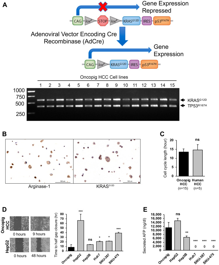
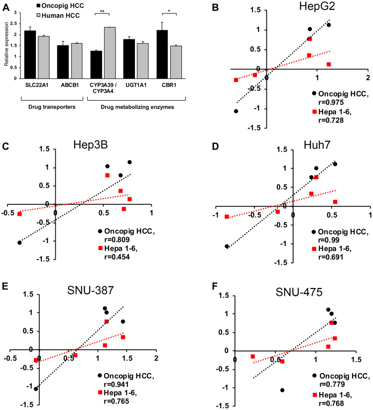
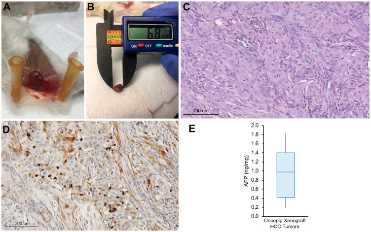
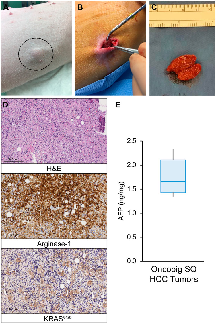
Hepatocellular carcinoma (HCC) is the second leading cause of cancer-related death worldwide. New animal models that faithfully recapitulate human HCC phenotypes are required to address unmet clinical needs and advance standard-of-care therapeutics. This study utilized the Oncopig Cancer Model to develop a translational porcine HCC model which can serve as a bridge between murine studies and human clinical practice. Reliable development of Oncopig HCC cell lines was demonstrated through hepatocyte isolation and Cre recombinase exposure across 15 Oncopigs. Oncopig and human HCC cell lines displayed similar cell cycle lengths, alpha-fetoprotein production, arginase-1 staining, chemosusceptibility, and drug metabolizing enzyme expression. The ability of Oncopig HCC cells to consistently produce tumors in vivo was confirmed via subcutaneous (SQ) injection into immunodeficient mice and Oncopigs. Reproducible development of intrahepatic tumors in an alcohol-induced fibrotic microenvironment was achieved via engraftment of SQ tumors into fibrotic Oncopig livers. Whole-genome sequencing demontrated intrahepatic tumor tissue resembled human HCC at the genomic level. Finally, Oncopig HCC cells are amenable to gene editing for development of personalized HCC tumors. This study provides a novel, clinically-relevant porcine HCC model which holds great promise for improving HCC outcomes through testing of novel therapeutic approaches to accelerate and enhance clinical trials.

Keywords: interventional radiology; large animal model; liver cancer; personalized medicine; transgenic pigs.


Conflict of interest statement

CONFLICTS OF INTEREST Ron C. Gaba receives research support from Guerbet USA LLC, Janssen Research & Development LLC, the United States Department of Defense, and the United States National Institutes of Health. F. Edward Boas is a co-founder of Claripacs, LLC. He received research funding (investigator-initiated) from Guerbet. He received research support (investigator-initiated) from GE. He received research supplies (investigator-initiated) from Bayer. He received a research grant and speaker fees from Society of Interventional Oncology, which were sponsored by Guerbet. He attended research meetings sponsored by Guerbet. He is an investor in Labdoor, Qventus, CloudMedx, Notable Labs, and Xgenomes. He is the inventor and assignee on US patent 8233586, and is an inventor on US provisional patent applications 62/754,139 and 62/817,116. Regina M. Schwind receives research support from Guerbet USA LLC and Janssen Research & Development LLC. Lawrence B. Schook research support from Guerbet USA LLC, Janssen Research & Development LLC, the United States Department of Defense, and the United States National Institutes of Health. Kyle M. Schachtschneider receives research support from Guerbet USA LLC, Janssen Research & Development LLC, and the United States National Institutes of Health. The remaining authors—Lobna Elkhadragy, Sulalita Chaki, Hanna H. Chen, Mohammed El-Kebir, Kelly D. Garcia, Eileena F. Giurini, Grace Guzman, Francesca V. LoBianco, Mario F. Neto, Jordan L. Newson, Aisha Qazi, Maureen Regan, Lauretta A. Rund, Matthew C. Stewart, Faith M. Thomas, Herbert E. Whiteley, and Jiaqi Wu—have no reported conflicts of interest.

Figures

Figure 1
Figure 1. Oncopig and human HCC in vitro phenotypes.
(A) Schematic of Oncopig transgene construct and agarose gel electrophoresis of RT-PCR products confirming Oncopig transgene (KRASG12D and TP53R167H) expression following exposure to AdCre. (B) Positive arginase-1 and KRASG12D staining (brown) of cultured Oncopig HCC cell lines (20×). (C) Oncopig and human HCC cell cycle lengths. (D) Representative cell migration images depicting faster gap closure in Oncopig compared to HepG2 and half gap closure rates for Oncopig (n = 15 cell lines) and human HCC cells. (E) AFP secretion from Oncopig (n = 15 cell lines) and human HCC cells. Huh7, SNU-387, and SNU475 are known non-AFP producing cell lines. ns = non-significant, *denotes p-value < 0.05, **denotes p-value ≤ 0.01, ***denotes p-value ≤ 0.0001.
Figure 2
Figure 2. Oncopig, human, and murine HCC in vitro chemotherapeutic susceptibility.
(A) Gene expression levels in Oncopig (n = 3 cell lines) and human HCC cells (HepG2, Huh7, and Hep3B). (BF) Correlation analysis of logIC50 values demonstrating more similar in vitro chemotherapeutic responses between Oncopig and human compared to murine Hepa1-6 and human HCC cells. Chemotherapeutic response of each HCC cell line towards sorafenib, doxorubicin, cisplatin, mitomycin C, and 5-FU was determined. Pearson correlation between logIC50 in Oncopig HCC cells or murine Hepa1-6 cells and the following human HCC cells was analyzed: (B) HepG2, (C) Hep3B, (D) Huh7, (E) SNU-387, and (F) SNU-475. *denotes P < 0.05, **denotes P ≤ 0.001.
Figure 3
Figure 3. Oncopig HCC xenograft tumor development.
(A) Representative SQ Oncopig HCC xenograft tumor. (B) Excised Oncopig HCC xenograft tumor. (C) H & E (20×) of Oncopig HCC xenograft tumor reveals densely cellular subcutaneous nodule with interspersed fat cells. Intervening fibrous vascular septae noted. (D) On arginase-1 IHC (20×), epithelial cells show focal arginase-1 expression (brown) consistent with hepatocellular differentiation. (E) AFP expression across Oncopig HCC xenograft tumors (n = 10).
Figure 4
Figure 4. Oncopig SQ HCC autograft formation.
(A) Photograph of visible SQ HCC tumor (circled) in Oncopig flank. (B) Excision of 2.0 cm SQ HCC tumor. (C) Excised and transected SQ HCC tumor. (D) H & E (20×) of Oncopig SQ HCC tumor demonstrates prominent, dispersed, pleomorphic large atypical cells, 5–10× the size of lymphocytes, flanking fibrous vascular septae, surrounded by dense mixed immune cell infiltrates. Arginase-1 IHC (20×) shows that these atypical cells show patchy arginase-1 expression (brown) consistent with hepatocellular differentiation. KRASG12D IHC (20×) confirms KRASG12D expression (brown) consistent with malignancy. (E) AFP expression across Oncopig SQ HCC tumors (n = 6).
Figure 5
Figure 5. Oncopig intrahepatic HCC tumor formation.
(A) Liver ultrasound depicting a hypoechoic 1 cm round intrahepatic HCC tumor (circled, L = liver, GB = gallbladder). (B) Contrast enhanced liver CT depicts same HCC tumor (circled). (C) Photograph of transected intrahepatic HCC tumor. (D) H & E (20×) of Oncopig intrahepatic HCC tumor reveals architectural distortion characterized by expansion of liver cords, nuclear pleomorphism, anisonucleosis, and nodular fibrosis. Masson’s trichrome of adjacent non-tumorous liver demonstrates dense collagen bands (arrows) consistent with METAVIR grade 2-3 fibrosis. (E) Arginase-1 IHC (20×) shows patchy arginase-1 expression (brown) consistent with hepatocellular differentiation. KRASG12D IHC (20×) confirms KRASG12D expression (brown) consistent with malignancy.
Figure 6
Figure 6. Genomic signatures of Oncopig HCC.
(A) Somatic copy-number calling reveals a largely copy-neutral tumor in line with the young age of the tumor. (B) Representative venn diagram showing distribution of SNVs in the cell line and 2 out of 5 tumor samples. (C) Mutational signatures identified resemble signatures observed in human HCC tumors (Signatures 1, 12, and 17).
Figure 7
Figure 7. CRISPR/Cas9-mediated disruption of Oncopig KRASG12D and TP53R167H transgenes.
(A) Schematic representation of the Oncopig transgene showing gRNA target sites and primers used for PCR. IRES, Internal ribosome entry site. (B) KRASG12D and TP53R167H editing efficiencies at multiple time points post transfection with Cas9 and gRNAs. (C) Frameshift mutations resulting in protein truncation for 2 Oncopig TP53R167H KO HCC cell lines developed via single cell clone isolation and screening. Dashed line marks the cleavage position, and dashed grey boxes represent nucleotide deletions. Dotted regions represent frameshifts in predicted protein sequences. (D) Positive arginase-1 staining (brown) of parental and TP53R167H KO cell lines (scale bar, 300 μm). (E) Cellular proliferation of Oncopig parental and TP53R167H KO HCC cell lines. Values represent mean ± S. D. (n ≥ 3). **indicates P < 0.001.

References

    1. Fitzmaurice C, Allen C, Barber RM, Barregard L, Bhutta ZA, Brenner H, Dicker DJ, Chimed-Orchir O, Dandona R, Dandona L, Fleming T, Forouzanfar MH, et al.; Global Burden of Disease Cancer Collaboration. Global, Regional, and National Cancer Incidence, Mortality, Years of Life Lost, Years Lived With Disability, and Disability-Adjusted Life-years for 32 Cancer Groups, 1990 to 2015: A Systematic Analysis for the Global Burden of Disease Study. JAMA Oncol. 2017; 3:524–548. 10.1001/jamaoncol.2016.5688. - DOI - PMC - PubMed
    1. Rahib L, Smith BD, Aizenberg R, Rosenzweig AB, Fleshman JM, Matrisian LM. Projecting cancer incidence and deaths to 2030: the unexpected burden of thyroid, liver, and pancreas cancers in the United States. Cancer Res. 2014; 74:2913–2921. 10.1158/0008-5472.CAN-14-0155. - DOI - PubMed
    1. Singal AG, El-Serag HB. Hepatocellular Carcinoma From Epidemiology to Prevention: Translating Knowledge into Practice. Clin Gastroenterol Hepatol. 2015; 13:2140–2151. 10.1016/j.cgh.2015.08.014. - DOI - PMC - PubMed
    1. National Cancer Institute Surveillance, Epidemiology, and End Results Program (NCI SEER). Cancer Stat Facts: Liver and Intrahepatic Bile Duct Cancer. 2019. Available from: https://seer.cancer.gov/statfacts/html/livibd.html.
    1. Moon AM, Singal AG, Tapper EB. Contemporary Epidemiology of Chronic Liver Disease and Cirrhosis. Clin Gastroenterol Hepatol. 2019; S1542–3565:30849–3. 10.1016/j.cgh.2019.07.060. - DOI - PMC - PubMed