

Conjunctival Sparing Ptosis Correction by White-Line Advancement Technique
- PMID: 32733700
- PMCID: PMC7378591
- DOI: 10.1155/2020/9021848
Conjunctival Sparing Ptosis Correction by White-Line Advancement Technique
Abstract
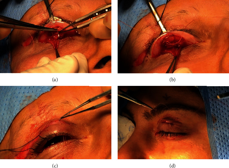
Purpose: To describe a modified technique of white line advancement posterior ptosis surgery and to report the success rate of the procedure.
Methods: A retrospective case series of 60 patients who presented with ptosis with good levator function. The success rate was defined as an MRD1 of greater than or equal to 3.5 mm, symmetrical eyelid position with an intereyelid height asymmetry of ≤1 mm, and a satisfactory eyelid contour at 3 months follow-up.
Results: Sixty patients (91 eyelids) met the inclusion criteria. Mild postoperative complications occurred in 11 patients that resolved without surgical intervention. Seven patients had recurrence of ptosis: four patients had early recurrence and 3 had late recurrence. The success rate was 88.33% with an average follow-up of 9 months.
Conclusion: This procedure is a promising technique in cosmetic and functional ptosis correction. The advantage of this posterior approach procedure is that there is no conjunctival resection; it is suitable for young patients who do not have excess eyelid skin. The procedure is quick with a short recovery period. Additionally, it can be combined with another procedure and in different pathology.
Copyright © 2020 Fatima A. Habroosh and Habibullah Eatamadi.
Conflict of interest statement
The authors declare that they have no conflicts of interest.
Figures


References
-
- Elbakary M. Posterior approach levator aponeurosis advancement in aponeurotic ptosis repair. Delta Journal of Ophthalmology. 2015;16(1):p. 32. doi: 10.4103/1110-9173.157787. - DOI
-
- Bowman W. Report of a chief operation performed at the royal london ophthalmic hospital for the quarter ending 25th of september 1857. The Royal London Ophthalmic Hospital Reports. 1859;11:p. 34.
-
- Blaskovicz L. A new operation for ptosis with shortening of the levator and tarsus. Archives of ophthalmology. 1923;52:563–573.
-
- Blaskovicz L. Treatment of ptosis. The formation of a fold in the eyelid and resection of the levator and tarsus. Archives of ophthalmology. 1929;1:672–680.
-
- Agatston S. A. Resection of Levator Palpebrae muscle by the conjunctival route for ptosis. Archives of Ophthalmology. 1942;27(5):994–996. doi: 10.1001/archopht.1942.00880050164011. - DOI
LinkOut - more resources
Full Text Sources
Medical
Miscellaneous
