. 2020 Jul 17;10(14):7512-7525.
doi: 10.1021/acscatal.0c01487. Epub 2020 Jun 8.

In Vivo Selection for Formate Dehydrogenases with High Efficiency and Specificity toward NADP

Affiliations

In Vivo Selection for Formate Dehydrogenases with High Efficiency and Specificity toward NADP

Liliana Calzadiaz-Ramirez et al. ACS Catal. .

Abstract

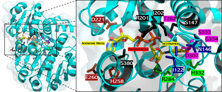
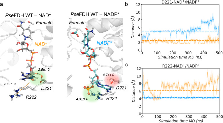
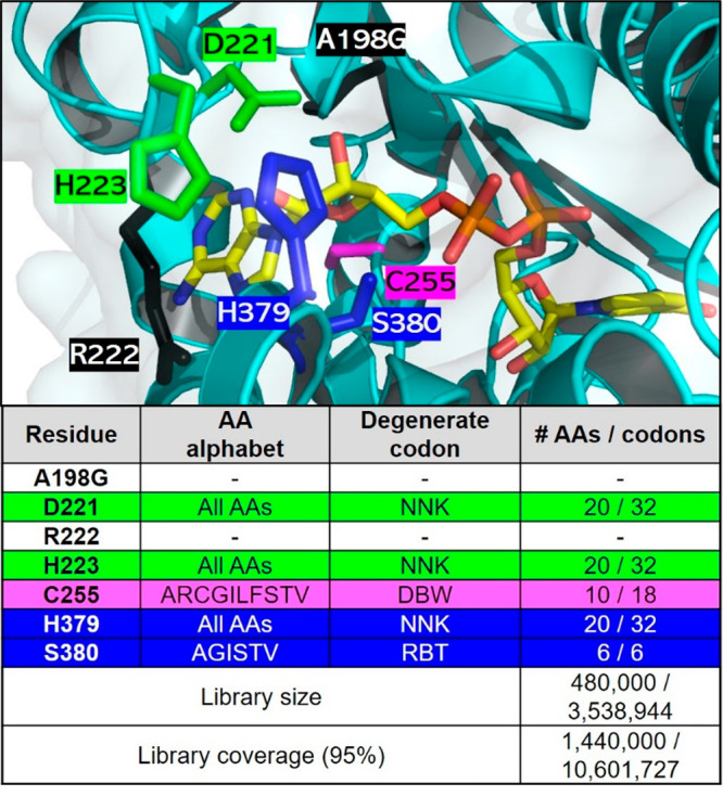
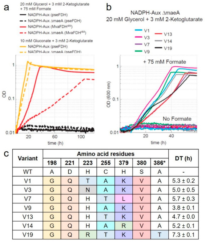
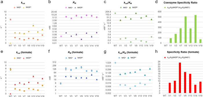
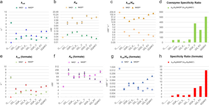
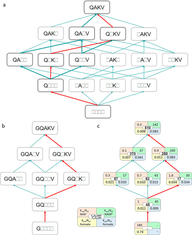
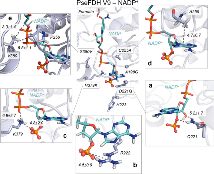
The efficient regeneration of cofactors is vital for the establishment of biocatalytic processes. Formate is an ideal electron donor for cofactor regeneration due to its general availability, low reduction potential, and benign byproduct (CO2). However, formate dehydrogenases (FDHs) are usually specific to NAD+, such that NADPH regeneration with formate is challenging. Previous studies reported naturally occurring FDHs or engineered FDHs that accept NADP+, but these enzymes show low kinetic efficiencies and specificities. Here, we harness the power of natural selection to engineer FDH variants to simultaneously optimize three properties: kinetic efficiency with NADP+, specificity toward NADP+, and affinity toward formate. By simultaneously mutating multiple residues of FDH from Pseudomonas sp. 101, which exhibits practically no activity toward NADP+, we generate a library of >106 variants. We introduce this library into an E. coli strain that cannot produce NADPH. By selecting for growth with formate as the sole NADPH source, we isolate several enzyme variants that support efficient NADPH regeneration. We find that the kinetically superior enzyme variant, harboring five mutations, has 5-fold higher efficiency and 14-fold higher specificity in comparison to the best enzyme previously engineered, while retaining high affinity toward formate. By using molecular dynamics simulations, we reveal the contribution of each mutation to the superior kinetics of this variant. We further determine how nonadditive epistatic effects improve multiple parameters simultaneously. Our work demonstrates the capacity of in vivo selection to identify highly proficient enzyme variants carrying multiple mutations which would be almost impossible to find using conventional screening methods.


Conflict of interest statement

The authors declare the following competing financial interest(s): A.B.-E. is a cofounder of b.fab, exploring the commercialization of microbial bioproduction using formate as a feedstock. The company was not involved in any way in performing or funding this study.

Figures

Figure 1
Figure 1
Active site of a PseFDH monomer. Catalytic residues involved in hydride transfer (green) and formate binding (blue) as well as those interacting with cofactor moieties of adenosine ribose (brown), phosphodiester (black), and nicotinamide (violet) are highlighted. Carbon (yellow), nitrogen (blue), and oxygen (red) atoms of the cofactor are shown in stick format. The picture was created using PyMol and PDB file 2NAD.
Figure 2
Figure 2
PseFDH WT NAD+/NADP+ conformational dynamics. (a) Representative structures of a PseFDH wild-type (WT) active site in the presence of NAD+ (gray, left) or NADP+ (cyan, right) and formate extracted from MD simulations (most populated clusters). The presence of the 2′- phosphate group of NADP+ causes a rearrangement of binding pocket residues. In WT-NAD+, the hydrogen bond interaction between D221 and the hydrogen of the 2′-OH group of NAD+ is highlighted in green. In WT-NADP+, the repulsive interaction between D221 and the 2′-phosphate group of NADP+ is shown in red and the salt-bridge interaction between R222 and the 2′-phosphate group of NADP+ in green. Relevant average distances (in Å) obtained from MD simulations are also depicted. (b) Plot of the distance between the carbon of the carboxylate group of D221 and either the 2′-OH group of NAD+ (orange) or the 2′-phosphate group of NADP+ (blue) along representative 500 ns replicas of MD simulations. Average distances (dashed orange line for WT-NAD+ and dashed blue line for WT-NADP+) of 2.5 ± 1.2 and 4.7 ± 1.0 Å are also shown, respectively. (c) Plot of the distance between the carbon of the guanidinium group of R222 and either the oxygen of the 2′-OH group of NAD+ (orange) or the 2′-phosphate group of NADP+ (blue) along representative 500 ns replicas of MD simulations. Average distances (dashed orange line for WT-NAD+ and dashed blue line for WT-NADP+) of 6.2 ± 1.9 and 4.3 ± 0.4 Å are also included, respectively. All distances are represented in Å. The trajectories of the three independent replicates are shown in Figure S2.
Figure 3
Figure 3
PseFDH CAST library design. (top) Active site of PseFDH variant A198G (black) highlighting residues selected for mutagenesis for switching cofactor specificity (green and violet), while recovering activity (blue). Residue R222 (black) was not mutated despite being suggested for mutagenesis by CRS-SALAD, as explained above. The carbon (yellow), nitrogen (blue), and oxygen (red) atoms of the cofactor are shown in stick format. The picture was generated with PyMol using PDB file 2NAD. (bottom) Amino acid (AA) alphabet, degenerate codon (N = A/T/G/C, K = T/G, D = A/G/T, B = C/G/T, W = A/T, R = A/G), and library size and coverage. The library size was calculated using the program CASTER 2.0.
Figure 4
Figure 4
In vivo characterization of FDH variants. (a) Growth profiles of NADPH auxotroph strain with five gene deletions expressing MvaFDH4M (red line) or PseFDH in the presence of formate (black line) or gluconate (yellow line) as a control. Additional deletion of maeA results in a more robust selection strain (NADPH-aux) that displays slower growth with MvaFDH4M (red dotted line) in the presence of formate, no growth with PseFDH in the presence of formate (black dotted lines), and full growth with PseFDH in the presence of gluconate (yellow dotted lines). (b) Growth profiles of NADPH-aux when the seven PseFDH variants isolated from the selection are expressed. Strains were cultured in 20 mM glycerol, 3 mM keto-glutarate, and 75 mM formate. (c) Sequencing results of PseFDH variants with doubling time (DT) ± standard error from triplicates.
Figure 5
Figure 5
Enzyme kinetics of FDH variants. Parameters reported for coenzyme turnover (a), affinity (b), catalytic efficiency (c), and specific ratio (d) under saturating amounts of formate as well as for formate turnover (e), affinity (f), catalytic efficiency (g), and specificity ratio (h) under saturating concentrations of coenzymes. The values represent average ± standard error from triplicates. Michaelis–Menten values and curves are found in Table S2 and Figure S16.
Figure 6
Figure 6
Enzyme kinetics of FDH deconvolutants. Parameters for coenzyme turnover (a), affinity (b), catalytic efficiency (c), and specific ratio (d) under saturating amounts of formate as well as for formate turnover (e), affinity (f), catalytic efficiency (g), and specificity ratio (h) under saturating concentrations of coenzymes. The values represent average ± standard error from triplicates. Michaelis–Menten values and curves are found in Table S3 and Figure S17.
Figure 7
Figure 7
Fitness landscapes in variants derived from PseFDH V9. (a) The introduction of the four additional mutations (D221Q, C255A, H379K, and S380V) into variant A198G yields 4! = 24 possible evolutionary pathways. Thick lines indicate the four possible pathways that can be explored by stepwise evolution with the available double and triple mutants. (b) Variants and the four possible pathways from double mutant A198G/D221Q (GQ) via triple variants A198G/D221Q/C255A (GQA), A198G/D221Q/S380V (GQV), and A198G/D221Q/H379K (GQK) and quadruple mutants A198G/D221Q/C255A/S380V (GQAV) and A198G/D221Q/H379K/S380V (GQKV) toward PseFDH V9 or quintuple mutant A198G/D221Q/C255A/H379K/S380V (GQAKV). (c) Catalytic efficiencies (mM–1 s–1) of the deconvoluted variants (shown in the left) toward either NAD+/formate (orange/yellow) or NADP+/formate (green/blue). The coenzyme specificity ratio (CSR) toward NADP+ ((kcat/KM)NADP+/(kcat/KM)NAD+) is shown in the middle (white). The red lines correspond to the pathway exemplified and described in the main text.
Figure 8
Figure 8
Conformational dynamics of PseFDH V9. A representative structure of the reshaped active site with NADP+ (cyan) and formate extracted from MD simulations (most populated cluster) is shown in the center with introduced mutations (Cα atoms depicted as spheres). (a) Representative structure of hydrogen bonds between D221Q and 2′-phosphate and the 3′-OH group of NADP+. The average distance between the amide group of D221Q and 3′-OH group of NADP+ (5.2 ± 1.7 Å) is depicted. (b) Representative structure of the salt-bridge interaction between the guanidinium group of R222 and the 2′-phosphate group of NADP+ (mean distance of 4.5 ± 0.9 Å) and the cation−π interaction between the guanidinium group of R222 and the adenine group of NADP+. (c) Representative structure of the salt-bridge interaction between the amino group of H379K and the 2′-phosphate group of NADP+ (4.8 ± 2.0 Å) and the salt-bridge interaction between the amino group of H379K and the linker 4′-phosphate group of NADP+ (6.9 ± 2.7 Å). (d) Representative structure of the CH−π interaction between the adenine ring of NADP+ and the β-carbon of the side chain of C255A. The average distance between the center of mass (COM) of the NADP+ adenine ring and the side chain of C255A (4.7 ± 0.7 Å) is depicted. (e) Representative structure of the interactions between the side chain of S380V and the side chain of P256 (with an average distance of 6.5 ± 1.1 Å) and the interactions between the side chain of S380V and the nicotinamide ribose group of NADP+ (8.3 ± 1.4 Å). All representative structures are extracted from the most populated clusters of three replicas of 500 ns of MD simulations for V9-NADP+. All distances are represented in Å.

References

    1. Claassens N. J.; Burgener S.; Vogeli B.; Erb T. J.; Bar-Even A. A critical comparison of cellular and cell-free bioproduction systems. Curr. Opin. Biotechnol. 2019, 60, 221–229. 10.1016/j.copbio.2019.05.003. - DOI - PMC - PubMed
    1. Hummel W.; Groger H. Strategies for regeneration of nicotinamide coenzymes emphasizing self-sufficient closed-loop recycling systems. J. Biotechnol. 2014, 191, 22–31. 10.1016/j.jbiotec.2014.07.449. - DOI - PubMed
    1. Andexer J. N.; Richter M. Emerging enzymes for ATP regeneration in biocatalytic processes. ChemBioChem 2015, 16, 380–386. 10.1002/cbic.201402550. - DOI - PubMed
    1. Babel W. The Auxiliary Substrate Concept: From simple considerations to heuristically valuable knowledge. Eng. Life Sci. 2009, 9, 285–290. 10.1002/elsc.200900027. - DOI
    1. Tishkov V. I.; Popov V. O. Protein engineering of formate dehydrogenase. Biomol. Eng. 2006, 23, 89–110. 10.1016/j.bioeng.2006.02.003. - DOI - PubMed