. 2020 Nov;26(11):1168-1177.
doi: 10.1111/cns.13441. Epub 2020 Jul 30.

Evaluation of a tumor electric field treatment system in a rat model of glioma

Affiliations

Evaluation of a tumor electric field treatment system in a rat model of glioma

Hao Wu et al. CNS Neurosci Ther. 2020 Nov.

Abstract

Objective: Glioma is a devastating disease lacking effective treatment. Tumor electric field therapy is emerging as a novel non-invasive therapy. The current study evaluates the efficacy and safety of a self-designed tumor electric field therapy system (TEFTS ASCLU-300) in a rat orthotopic transplantation model of glioma.

Methods: A model of intracranial orthotopic transplantation was established in rats using glioma C6 cells. For electric field therapy, glioma-bearing rats were exposed to alternating electric fields generated by a self-developed TEFTS starting on either 1st (Group 2) or 3rd (Group 3) day after transplantation, while other conditions were maintained the same as non-treated rats (Group 1). Glioma size, body weight, and overall survival (OS) were compared between groups. Immunohistochemical staining was applied to access tumor cell death and microvessel density within the tumor. In addition, the systemic effects of TEFTS on blood cells, vital organs, and hepatorenal functions were evaluated.

Results: TEFTS treatment significantly elongated the OS of tumor-bearing rats compared with non-treated rats (non-treated vs treated: 24.77 ± 7.08 days vs 40.31 ± 19.11 days, P = .0031). Continuous TEFTS treatment starting on 1st or 3rd day significantly reduced glioma size at 2 and 3 weeks after tumor cell inoculation (Week 2: Group 1:289.95 ± 101.69 mm3 ; Group 2:70.45 ± 17.79 mm3 ; Group 3:73.88 ± 33.21 mm3 , P < .0001. Week 3: Group 1:544.096 ± 78.53 mm3 ; Group 2:187.58 ± 78.44 mm3 ; Group 3:167.14 ± 109.96 mm3 , P = .0005). Continuous treatment for more than 4 weeks inhibited tumor growth. The TEFTS treatment promoted tumor cell death, as demonstrated by increased number of Caspase 3+ cells within the tumor (non-treated vs treated: 38.06 ± 10.04 vs 68.57 ± 8.09 cells/field, P = .0007), but had minimal effect on microvessel density, as shown by CD31 expression (non-treated vs treated: 1.63 ± 0.09 vs 1.57 ± 0.13% of positively stained areas, P > .05). No remarkable differences were observed in hepatorenal function, blood cell counts, or other vital organs between non-treated and treated groups.

Conclusion: The TEFTS developed by our research team was proved to be effective and safe to inhibit tumor growth and improve general outcomes in a rat model of brain glioma.

Keywords: cell death; electric field therapy; glioma; survival; tumor electric field treatment system; tumor size.


Conflict of interest statement

The authors declare no conflict of interest.

Figures

FIGURE 1
FIGURE 1
Flow chart of experimental design and main endpoint measurements
FIGURE 2
FIGURE 2
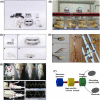
Tumor Electric Field Treatment System and Operation. A, Tumor Electric Field Treatment System (TEFTS, ASCLU‐300). B, Instrument operation during experiments. C, Main components of TEFTS. a, Battery and charger; b, Junction box and connecting cables; c, the electric field generator; d, Electrodes. D, Electrodes and accessories. a, On the back of electrode; b, Thermistor and circuit; c, after installing thermistor and circuit; d, Two pairs of ceramic disk electrodes. Thermistors on the back of electrodes are connected to a temperature‐controlling device. The diameter of the disk electrode is 8 mm (dielectric constant ε ≈ 10 000, capacitance c ≈ 10 nF). Four electrodes are used as one treatment unit. The wire is coated with metal to prevent from gnawing; e, two positive‐negative pairs of electrodes connected by a flexible circuit board are arranged perpendicularly to each other. The surface of the electrode is covered with hydrogel, and the back is fixed on medical adhesive tapes (1. Thermistor; 2. Disk ceramic electrode; 3. Metal piping coated wires; 4. Flexible circuit board; 5. Medical adhesive tape). E, Installation of electrodes and measurement of electric fields. a, the red dots indicate the locations of four electrodes on the head of a rat; b, the electrodes are fixed on head by tapes; c, a head cover protects the electrode from being scratched by the rat with its forelegs; d, the electrodes and a head cover are fixed; e, f, the voltage measured from the scalp; g, h, the intracranial voltage measured. F, Schematic diagram showing the generation of alternating electric field. Waveform generator built in the field programmable gate array (FPGA) generates the digital signal of sine wave according to the preset parameters. The digital signal is converted into analog signal by a high‐speed discretionary access control (DAC) module, and then filtered. The analog signal is amplified by a power amplifier, outputs to the electrode plate affixed to the therapeutic target, and generates an alternating electric field inside the target
FIGURE 3
FIGURE 3
Effect of TEFTS on glioma growth in rats. A, Body weight. B, Survival curve. C, Tumor growth over time was monitored by MRI. Representative MRI images of TEFTS‐treated rats and non‐treated control rats from 1st to 8th weeks after tumor transplantation (a). Continuous TEFTS treatment significantly reduced tumor volume at 2nd and 3rd week after tumor inoculation (b‐d). Changes of tumor volume over time in individual rat were displayed in (e). D, Continuous TEFTS treatment for 8 wk. In the first 4 wk, the tumor volume gradually increased. The tumor volume was significantly reduced from the 5th week to the 8th week. After stopping the treatment, no rebound was observed at the 10th and 12th week. The white arrows show the tumor, orange arrows indicate the area after the tumor shrinks/disappears. *P < .05;** P < .01; ***P < .001; ****P < 0001
FIGURE 4
FIGURE 4
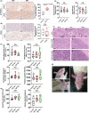
Safety of the TEFTS. A, Immunohistochemical staining of cleaved caspase‐3. The number of positive cells was counted under microscope (40×). B, Immunohistochemical staining of CD31. The area of positive staining was quantified. C‐E, Blood routine examination in each group on the 15th day of treatment. C, Blood cell counts; (D) alanine aminotransferase (ALT) and aspartate aminotransferase (AST) levels; (E), Urea nitrogen and serum creatinine levels among the three groups. F, Immunohistochemical staining of brain, liver, and kidney slices collected from the rats in different groups. G, Mild contact dermatitis on the skin where the electrode is attached. a,b, Mild rash in different parts of the electrode contacted areas; c, One week after the use of corticosteroids, the rash obviously has disappeared. *P < .05;**P < 01; ***P < .001; ****P < .0001. Scale bar = 100 μm

References

    1. GBD 2016 Brain and Other CNS Cancer Collaborators . Global, regional, and national burden of brain and other CNS cancer, 1990–2016: a systematic analysis for the Global Burden of Disease Study. Lancet Neurol. 2019;18(4):376‐393. - PMC - PubMed
    1. Ostrom QT, Cote DJ, Ascha M, Kruchko C, Barnholtz‐Sloan JS. Adult glioma incidence and survival by race or ethnicity in the United States from 2000 to 2014. JAMA Oncol. 2018;4(9):1254‐1262. - PMC - PubMed
    1. Delgado‐Martin B, Medina MA. Advances in the knowledge of the molecular biology of glioblastoma and its impact in patient diagnosis, stratification, and treatment. Adv Sci (Weinh). 2020;7(9):1902971. - PMC - PubMed
    1. Chongsathidkiet P, Jackson C, Koyama S, et al. Sequestration of T cells in bone marrow in the setting of glioblastoma and other intracranial tumors. Nat Med. 2018;24(9):1459‐1468. - PMC - PubMed
    1. Wenger C, Miranda PC, Salvador R, et al. A review on tumor‐treating fields (TTFields): clinical implications inferred from computational modeling. IEEE Rev Biomed Eng. 2018;11:195‐207. - PubMed