Case Reports
. 2020 Nov;102(9):e1-e4.
doi: 10.1308/rcsann.2020.0137. Epub 2020 Jul 31.

Extra-abdominal manifestations of retroperitoneal infection: a case of popliteal sinus secondary to duodenal ulcer

Affiliations
Case Reports

Extra-abdominal manifestations of retroperitoneal infection: a case of popliteal sinus secondary to duodenal ulcer

L Granger et al. Ann R Coll Surg Engl. 2020 Nov.

Abstract

Retroperitoneal abscesses can be gastrointestinal, urological or vascular in origin, and can spread via the retrofascial compartment through the psoas muscle to the lower limb. We describe the case of a 73-year-old woman with right knee pain for three weeks, a cellulitic right thigh and cholestatic liver function tests. A purulent sinus developed in the popliteal fossa and computed tomography of the abdomen revealed a right-sided retroperitoneal collection with gas, extending to the right pelvis and inguinal region. The popliteal fossa sinus and retroperitoneal collection were identified as a single pathology through computed tomography, magnetic resonance imaging and culture of identical organisms. At laparotomy, perforated duodenal ulcer disease was identified as the cause of the retroperitoneal abscess. Clinicians should seek to exclude retroperitoneal sources of infection in cases of lower leg infection, including perforated duodenal ulcer, caecal adenocarcinoma and appendicitis.

Keywords: Abdominal abscess; Duodenal ulcer surgery; Lower extremity surgery; Retroperitoneal space.


Figures

Figure 1
Figure 1
Axial computed tomography of the abdomen with contrast at L4 level showing an abscess in the right paracolic gutter in retroperitoneum with gas
Figure 2
Figure 2
Coronal computed tomography of the abdomen and pelvis with contrast; gas and abscess visualised in the right paracolic gutter in retroperitoneum
Figure 3
Figure 3
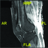
Sagittal magnetic resonance imaging of the femur with gadolinium contrast showing subcutaneous oedema in right thigh and collection in posterior thigh
Figure 4
Figure 4
Axial magnetic resonance imaging of the femur with gadolinium contrast showing subcutaneous oedema and collection in right posterior thigh
Figure 5
Figure 5
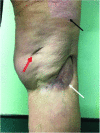
Posterior view of the knee at one-year follow-up. Donor site of split skin graft (white arrow). Site of initial discharging sinus and resultant recipient graft site (black arrow). Healed site of a second sinus (red arrow).

References

    1. Simons G, Sty J, Starshak R. Retroperitoneal and retrofascial abscesses: a review. J Bone Joint Surg 1983; : 1041–1058. - PubMed
    1. Mallia A, Ashwood N, Arealis G, Galanopoulos I. Retroperitoneal abscess: an extra-abdominal manifestation. BMJ Case Rep 2015; : bcr2014207437. - PMC - PubMed
    1. Das A, Hureibi K, Tayyab M, McCullough P. Caecal cancer presenting as leg erythema and a lung opacity. BMJ Case Rep 2017; : bcr2017220752. - PMC - PubMed
    1. Timbrell S, Straiton D, Breaks E et al. . Necrotising soft tissue infection of the lower limb due to a perforated caecal carcinoma: a case report. J Med Case Rep 2014; : 80. - PMC - PubMed
    1. Hsieh C, Wang Y, Yang H et al. . Extensive retroperitoneal and right thigh abscess in a patient with ruptured retrocecal appendicitis: an extremely fulminant form of a common disease. World J Gastroenterol 2006; : 496. - PMC - PubMed

Publication types

MeSH terms