Review
. 2020 Jan-Dec:26:1076029620943293.
doi: 10.1177/1076029620943293.

COVID-19-Associated Coagulopathy: An Exacerbated Immunothrombosis Response

Affiliations
Review

COVID-19-Associated Coagulopathy: An Exacerbated Immunothrombosis Response

Apoorva Jayarangaiah et al. Clin Appl Thromb Hemost. 2020 Jan-Dec.

Abstract

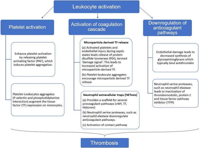
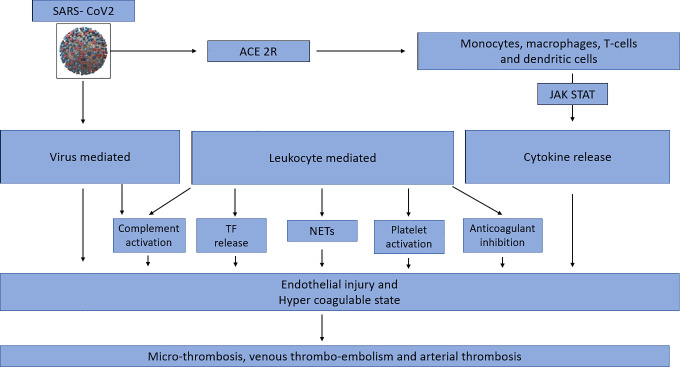
Since the onset of the global pandemic in early 2020, coronavirus disease 2019 (COVID-19) has posed a multitude of challenges to health care systems worldwide. In order to combat these challenges and devise appropriate therapeutic strategies, it becomes of paramount importance to elucidate the pathophysiology of this illness. Coronavirus disease 2019, caused by the novel severe acute respiratory syndrome coronavirus 2 (SARS-CoV2), is characterized by a dysregulated immune system and hypercoagulability. COVID-associated coagulopathy (CAC) was recognized based on profound d-dimer elevations and evidence of microthrombi and macrothrombi, both in venous and arterial systems. The underlying mechanisms associated with CAC have been suggested, but not clearly defined. The model of immunothrombosis illustrates the elaborate crosstalk between the innate immune system and coagulation. The rendering of a procoagulant state in COVID-19 involves the interplay of many innate immune pathways. The SARS-CoV2 virus can directly infect immune and endothelial cells, leading to endothelial injury and dysregulation of the immune system. Activated leukocytes potentiate a procoagulant state via release of intravascular tissue factor, platelet activation, NETosis, and inhibition of anticoagulant mechanisms. Additional pathways of specific relevance in CAC include cytokine release and complement activation. All these mechanisms have recently been reported in COVID-19. Immunothrombosis provides a comprehensive perspective of the several synergistic pathways pertinent to the pathogenesis of CAC.

Keywords: COVID-19; coagulopathy; immunothrombosis.


Conflict of interest statement

Declaration of Conflicting Interests: The author(s) declared no potential conflicts of interest with respect to the research, authorship, and/or publication of this article.

Figures

Figure 1.
Figure 1.
Overview of mechanisms of hemostasis. The coagulation cascade during hemostasis is initiated with the release of tissue factor (TF). (1) At the site of endothelial injury, TF that is stored in the adventitial and medial layers of the vessel wall is released. Platelet activation and adhesion occurs at the site of injury. (1) Encrypted TF is also released from monocyte-derived microparticles. Encrypted TF is activated through the secretion of protein disulfide isomerase (PDI), which occurs because of endothelial injury and platelet activation. (2) Activated TF will form a complex with circulating factor VIIa (FVIIa), leading to activation of FX to FXa. A small amount of thrombin generated creates an amplification loop by activating FVIII, FXI, and FV. (3) During the propagation phase, FIXa, which is generated by TF–VIIa complex or FXIa, binds to its cofactor FVIIIa to form the intrinsic tenase complex. FXa is then formed by TF–FVIIa complex or FIXa–FVIIa complex. (4) FXa binds to FVa, forming the prothrombinase complex. (5) FXa–FVa complex converts prothrombin to thrombin. (6) Thrombin further activates fibrinogen to fibrin, which is cross-linked by FXIIIa to form a stable clot.
Figure 2.
Figure 2.
Principles of immunothrombosis. Monocytes and their microvesicles release activated intravascular tissue factor (TF) in response to pathogen-associated molecular patterns (PAMPs) or damage-associated molecular patterns (DAMPs), which initiates the extrinsic pathway of coagulation. Activated platelets and endothelium secrete protein disulfide isomerase (PDI), which activates microvesicle-derived TF. Interaction between P-selectin on activated platelets and P-selectin glycoprotein ligand (PSGL) on leukocytes enhance recruitment of leukocytes and increase TF production. Neutrophil extracellular traps (NETs) provide a scaffold consisting of DNA, histones, and neutrophil serine proteases, and they support immunothrombosis through several pathways. (1) NETs bind to von Willebrand factor (VWF) and support the recruitment of platelets. (2) Histones H3 and H4 on NETs can trigger the activation of platelets. (3) NETs bind to TF leading to TF-mediated thrombin generation through the extrinsic pathway (dotted arrow). (4) Neutrophil elastase and other neutrophil serine proteases cleave and inactivate anticoagulants, including TF pathway inhibitor (TFPI) and thrombomodulin. (5) NETs can directly activate factor XII (the contact pathway of coagulation) mediated by platelet-derived polyphosphates (PolyP). The complement system (specifically the activated complement components C3a and C5a) also enhances platelet activation.
Figure 3.
Figure 3.
Summary of the mechanisms of leukocyte induced procoagulant state. Activation of leukocytes during states of sepsis and inflammation leads to a prothrombotic state, primarily through platelet activation, activation of coagulation cascade, and downregulation of anticoagulant pathways.
Figure 4.
Figure 4.
Model of COVID-associated coagulopathy. A, First, SARS-CoV2 gains entry into cells via the angiotensin-converting enzyme 2 receptor (ACE-2R), leading to activation of the innate immune system. Direct viral infection of the immune cells leads to dysregulation and cytokine release. B, Activated monocytes, macrophages stimulate Janus Kinas (JAK)- Signal transducer and activator of transcription (STAT) (JAK STAT) pathways via cis/trans signaling, leading to an amplification of cytokine release. C, SARS-CoV2 infects endothelial cells directly, resulting in endothelial injury and therefore propagating a hypercoagulable state. D, Leukocyte activation and ensuing cytokine storm in SARS-CoV2 leads to a hypercoagulable state though 5 main ways: increased tissue factor release, neutrophil extracellular traps, platelet activation, inactivation of anticoagulant pathways, and complement activation. E, Dysregulation of the immune system in SARS-CoV2 infection and the resultant endothelial injury and hypercoagulable state culminates into widespread microthrombosis, venous thromboembolism, and arterial thrombosis.

References

    1. Munster VJ, Koopmans M, Doremalen NV, Riel DV, Wit ED. A novel coronavirus emerging in China—key questions for impact assessment. N Eng J Med. 2020;382(8):692–694. doi:10.1056/nejmp2000929 - PubMed
    1. Zhou F, Yu T, Du R, et al. Clinical course and risk factors for mortality of adult inpatients with COVID-19 in Wuhan, China: a retrospective cohort study. Lancet. 2020;395(10229):1054–1062. doi:10.1016/s0140-6736(20)30566-3 - PMC - PubMed
    1. Engelmann B, Massberg S. Thrombosis as an intravascular effector of innate immunity. Nat Rev Immunol. 2012;13(1):34–45. doi:10.1038/nri3345 - PubMed
    1. Wang D, Hu B, Hu C, et al. Clinical characteristics of 138 hospitalized patients with 2019 novel coronavirus–infected pneumonia in Wuhan, China. JAMA. 2020;323(11):1061 doi:10.1001/jama.2020.1585 - PMC - PubMed
    1. Chen N, Zhou M, Dong X, et al. Epidemiological and clinical characteristics of 99 cases of 2019 novel coronavirus pneumonia in Wuhan, China: a descriptive study. Lancet. 2020;395(10223):507–513. doi:10.1016/s0140-6736(20)30211-7 - PMC - PubMed

MeSH terms