Comparative Study
. 2020 Jul 31;18(7):e3000811.
doi: 10.1371/journal.pbio.3000811. eCollection 2020 Jul.

Zinc protection of fertilized eggs is an ancient feature of sexual reproduction in animals

Affiliations
Comparative Study

Zinc protection of fertilized eggs is an ancient feature of sexual reproduction in animals

Katherine L Wozniak et al. PLoS Biol. .

Abstract

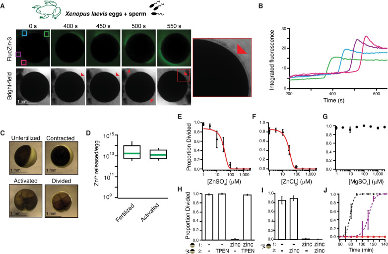
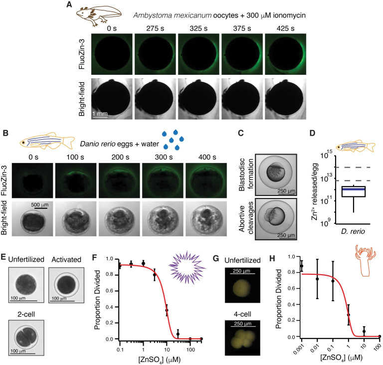
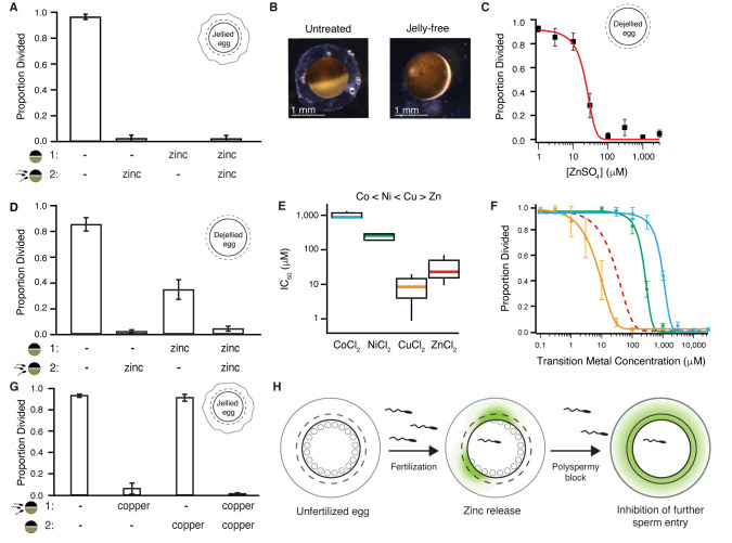
One of the earliest and most prevalent barriers to successful reproduction is polyspermy, or fertilization of an egg by multiple sperm. To prevent these supernumerary fertilizations, eggs have evolved multiple mechanisms. It has recently been proposed that zinc released by mammalian eggs at fertilization may block additional sperm from entering. Here, we demonstrate that eggs from amphibia and teleost fish also release zinc. Using Xenopus laevis as a model, we document that zinc reversibly blocks fertilization. Finally, we demonstrate that extracellular zinc similarly disrupts early embryonic development in eggs from diverse phyla, including Cnidaria, Echinodermata, and Chordata. Our study reveals that a fundamental strategy protecting human eggs from fertilization by multiple sperm may have evolved more than 650 million years ago.


Conflict of interest statement

The authors have declared that no competing interests exist.

Figures

Fig 1
Fig 1. X. laevis eggs release zinc at fertilization, which protects eggs from additional fertilizations.
(A) Representative fluorescence and bright-field images of X. laevis eggs in FluoZin-3 before and after sperm application. Time is relative to sperm addition. Zinc release coincided with lifting of the envelope (red arrowheads and insert) (N = 16 eggs, 4 trials). (B) Integrated fluorescence relative to time of sperm addition and detected by region of interest analysis, indicated by colored boxes in panel (A). (C) Representative images of X. laevis eggs and embryos. (D) Box plot distribution of zinc ions released per X. laevis egg upon fertilization or activation with 10 μM ionomycin as detected by FluoZin-3 fluorometry. (E-G) Proportion of inseminated eggs that developed cleavage furrows in indicated concentrations of ZnSO4, ZnCl2, or MgSO4. Plots in (A) and (B) were fit with sigmoidal functions. (H) Proportion of development of eggs subjected to a 15-minute pretreatment with 0 or 300 μM ZnSO4 (solution 1) then washed and moved to solution (2) with 0 or 300 μM TPEN for sperm addition (N = 68–259 eggs, 5 trials). (I) Incidence of cleavage furrow development from eggs inseminated in 0 or 1 mM ZnSO4 and then transferred to a new solution with 0 or 1 mM ZnSO4 30 minutes after sperm addition (N = 52–258 eggs in 6 trials). (J) Proportion of cleavage furrow development from eggs inseminated in and transferred to control conditions (black), eggs inseminated in 300 μM ZnSO4 and transferred to 600 μM TPEN 30 minutes following sperm addition (purple), or eggs inseminated in and transferred to 1 mM ZnSO4 (red) (N = 26–58 eggs in 7 trials). (E-J) Errors are SEM. For full source data, see S1 Data. TPEN, N,N,N′,N′-tetrakis(2-pyridinylmethyl)-1,2-ethanediamine.
Fig 2
Fig 2. Zinc release and protection of eggs is shared in external fertilizers.
(A) Representative FluoZin-3 and bright-field images of A. mexicanum oocytes before and after activation with 300 μM ionomycin (N = 6 oocytes, 2 trials). (B) Representative images of FluoZin-3 and bright-field of D. rerio eggs releasing zinc upon activation with water (N = 7 eggs, 4 trials). Eggs were in 50 μM FluoZin-3 (A and B). (C) Appearance of blastodisc and abortive cleavages confirmed activation for D. rerio eggs. (D) Box plot of zinc released per D. rerio egg upon activation as detected by fluorometry using FluoZin-3 (N = 59–138 eggs, 5 trials). Dashed lines denote the 25th and 75th percentile of the data distribution for zinc released from X. laevis eggs following fertilization. (E) Representative images of S. purpuratus eggs and embryos. (F) Development was blocked in a concentration-dependent manner for S. purpuratus eggs in ZnSO4 (N = 120–684 eggs, 5 trials). (G) Representative images of H. symbiolongicarpus unfertilized and divided eggs. (H) Development was blocked in a concentration-dependent manner for H. symbiolongicarpus eggs inseminated in extracellular ZnSO4 (N = 143–1463 eggs, 5–6 trials). Errors are SEM. For full source data, see S1 Data.
Fig 3
Fig 3. Zinc inhibits embryonic development by binding to proteins surrounding the egg.
(A) Incidence of development of eggs pretreated in 300 μM ZnSO4 prior to insemination (N = 34–150 eggs, 5 trials). (B) X. laevis eggs before and after jelly removal. (C) Incidence of development of jelly-free eggs in varying ZnSO4 concentrations (N = 53–96 eggs, 6 trials). (D) Average proportion of cleavage furrow development from de-jellied eggs pretreated in solution (1), with either 0 or 300 μM ZnSO4. After a 15-minute treatment, eggs were then washed and moved to solution (2) for sperm addition with 0 or 300 μM ZnSO4 (N = 35–192, 5 trials). (E) Box plot distributions of the IC50 values of inhibition of appearance of cleavage furrows in X. laevis eggs inseminated in cobalt (blue), nickel (green), copper (orange), or zinc (red). (F) Proportion of division of X. laevis eggs inseminated in varying concentrations of extracellular cobalt (N = 148–295 eggs, 5 trials), nickel (N = 136–283 eggs, 5 trials), or copper (N = 230–321 eggs, 5 trials). (G) Incidence of cleavage furrow development from eggs inseminated in solution (1), with 0 or 100 μM CuCl2, and then transferred to solution (2), with 0 or 100 μM CuCl2, 30 minutes after sperm addition (N = 205–302 eggs, 5 trials). (H) Fertilization stimulates the release of zinc from the egg to modify the envelope and jelly coat to inhibit sperm entry in X. laevis. All errors are SEM. For full source data, see S1 Data. IC50, half-maximal inhibitory concentration.

References

    1. Wong JL, Wessel GM. Defending the zygote: search for the ancestral animal block to polyspermy. Curr Top Dev Biol. 2006;72:1–151. 10.1016/S0070-2153(05)72001-9 . - DOI - PubMed
    1. Jaffe LA. Fast block to polyspermy in sea urchin eggs is electrically mediated. Nature. 1976;261(5555):68–71. Epub 1976/05/06. 10.1038/261068a0 . - DOI - PubMed
    1. Wozniak KL, Phelps WA, Tembo M, Lee MT, Carlson AE. The TMEM16A channel mediates the fast polyspermy block in Xenopus laevis. J Gen Physiol. 2018. Epub 2018/07/18. 10.1085/jgp.201812071 . - DOI - PMC - PubMed
    1. Wozniak KL, Tembo M, Phelps WA, Lee MT, Carlson AE. PLC and IP3-evoked Ca(2+) release initiate the fast block to polyspermy in Xenopus laevis eggs. J Gen Physiol. 2018. Epub 2018/07/18. 10.1085/jgp.201812069 . - DOI - PMC - PubMed
    1. Wozniak KL, Carlson AE. Ion channels and signaling pathways used in the fast polyspermy block. Mol Reprod Dev. 2020;87(3):350–7. Epub 2019/05/16. 10.1002/mrd.23168 - DOI - PMC - PubMed

Publication types

LinkOut - more resources