

Shear-dependent platelet aggregation size
- PMID: 32735693
- PMCID: PMC7818454
- DOI: 10.1111/aor.13783
Shear-dependent platelet aggregation size
Abstract
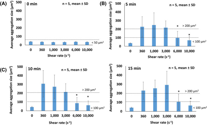
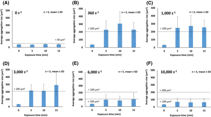
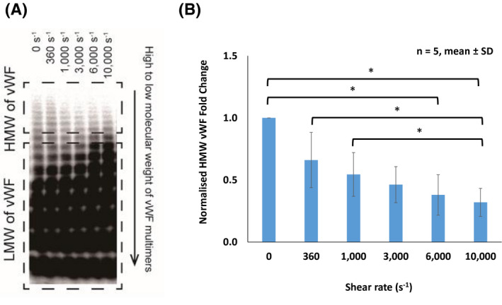
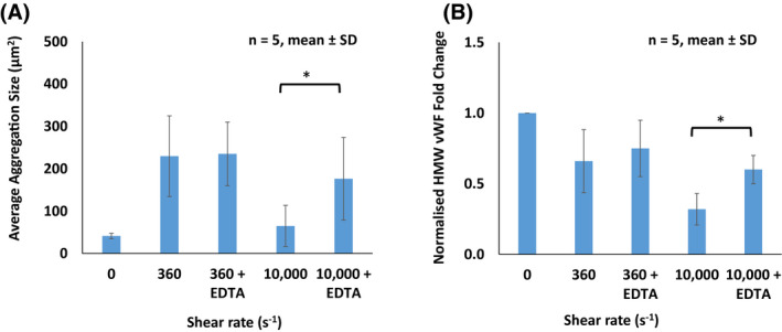
Nonsurgical bleeding is the most frequent complication of left ventricular assist device (LVAD) support. Supraphysiologic shear rates generated in LVAD causes impaired platelet aggregation, which increases the risk of bleeding. The effect of shear rate on the formation size of platelet aggregates has never been reported experimentally, although platelet aggregation size can be considered to be directly relevant to bleeding complications. Therefore, this study investigated the impact of shear rate and exposure time on the formation size of platelet aggregates, which is vital in predicting bleeding in patients with an LVAD. Human platelet-poor plasma (containing von Willebrand factor, vWF) and fluorochrome-labeled platelets were subjected to a range of shear rates (0-10 000 s-1 ) for 0, 5, 10, and 15 minutes using a custom-built blood-shearing device. Formed sizes of platelet aggregates under a range of shear-controlled environment were visualized and measured using microscopy. The loss of high molecular weight (HMW) vWF multimers was quantified using gel electrophoresis and immunoblotting. An inhibition study was also performed to investigate the reduction in platelet aggregation size and HMW vWF multimers caused by either mechanical shear or enzymatic (a disintegrin and metalloproteinase with a thrombospondin type 1 motif, member 13-ADAMTS13, the von Willebrand factor protease) mechanism under low and high shear conditions (360 and 10 000 s-1 ). We found that the average size of platelet aggregates formed under physiological shear rates of 360-3000 s-1 (200-300 μm2 ) was significantly larger compared to those sheared at >6000 s-1 (50-100 μm2 ). Furthermore, HMW vWF multimers were reduced with increased shear rates. The inhibition study revealed that the reduction in platelet aggregation size and HWM vWF multimers were mainly associated with ADAMTS13. In conclusion, the threshold of shear rate must not exceed >6000 s-1 in order to maintain the optimal size of platelet aggregates to "plug off" the injury site and stop bleeding.
Keywords: ADAMTS13; exposure time; platelet aggregate; shear rate; von Willebrand factor.
© 2020 The Authors. Artificial Organs published by International Center for Artificial Organ and Transplantation (ICAOT) and Wiley Periodicals LLC.
Conflict of interest statement
The authors declare that they have no conflicts of interest with the contents of this article.
Figures
References
-
- McMurray JJ, Adamopoulos S, Anker SD, Auricchio A, Bohm M, Dickstein K, et al. ESC Guidelines for the diagnosis and treatment of acute and chronic heart failure 2012: the Task Force for the Diagnosis and Treatment of Acute and Chronic Heart Failure 2012 of the European Society of Cardiology. Developed in collaboration with the Heart Failure Association (HFA) of the ESC. Eur Heart J. 2012;33:1787–847. - PubMed
-
- Slaughter MS, Rogers JG, Milano CA, Russell SD, Conte JV, Feldman D, et al. Advanced heart failure treated with continuous‐flow left ventricular assist device. N Eng J Med. 2009;361:2241–51. - PubMed
-
- Proudfoot AG, Davidson SJ, Strueber M. von Willebrand factor disruption and continuous‐flow circulatory devices. J Heart Lung Transplant. 2017;36:1155–63. - PubMed
-
- Katz JN, Adamson RM, John R, Tatooles A, Sundareswaran K, Kallel F, et al. Safety of reduced anti‐thrombotic strategies in HeartMate II patients: a one‐year analysis of the US‐TRACE study. J Heart Lung Transplant. 2015;34:1542–8. - PubMed
MeSH terms
Substances
Grants and funding
LinkOut - more resources
Full Text Sources
Miscellaneous
