. 2020 Sep 21;30(18):3664-3671.e4.
doi: 10.1016/j.cub.2020.06.090. Epub 2020 Jul 30.

The Tail of Kinesin-14a in Giardia Is a Dual Regulator of Motility

Affiliations

The Tail of Kinesin-14a in Giardia Is a Dual Regulator of Motility

Kuo-Fu Tseng et al. Curr Biol. .

Abstract

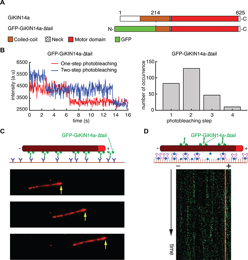
Kinesin-14s are microtubule-based motor proteins that play important roles in mitotic spindle assembly [1]. Ncd-type kinesin-14s are a subset of kinesin-14 motors that exist as homodimers with an N-terminal microtubule-binding tail, a coiled-coil central stalk (central stalk), a neck, and two identical C-terminal motor domains. To date, no Ncd-type kinesin-14 has been found to naturally exhibit long-distance minus-end-directed processive motility on single microtubules as individual homodimers. Here, we show that GiKIN14a from Giardia intestinalis [2] is an unconventional Ncd-type kinesin-14 that uses its N-terminal microtubule-binding tail to achieve minus-end-directed processivity on single microtubules over micrometer distances as a homodimer. We further find that although truncation of the N-terminal tail greatly reduces GiKIN14a processivity, the resulting tailless construct GiKIN14a-Δtail is still a minimally processive motor and moves its center of mass via discrete 8-nm steps on the microtubule. In addition, full-length GiKIN14a has significantly higher stepping and ATP hydrolysis rates than does GiKIN14a-Δtail. Inserting a flexible polypeptide linker into the central stalk of full-length GiKIN14a nearly reduces its ATP hydrolysis rate to that of GiKIN14a-Δtail. Collectively, our results reveal that the N-terminal tail of GiKIN14a is a de facto dual regulator of motility and reinforce the notion of the central stalk as a key mechanical determinant of kinesin-14 motility [3].

Keywords: TIRF microscopy; central stalk; dark-field microscopy; kinesin-14; microtubules; processivity; stepping.


Conflict of interest statement

Declaration of Interests The authors declare no competing interests.

Figures

Figure 1.
Figure 1.. Full-Length GiKIN14a Exhibits Minus-End-Directed Processivity over Micrometer Distances on Single Microtubules as Individual Homodimers.
(A) Schematic diagrams of full-length GiKIN14a, GFP-GiKIN14a-tail, and GFP-GiKIN14a. (B) Predicted coiled-coil profiles of the central stalks of GiKIN14a (red), Ncd-3xGS (blue), and Ncd-WT (dark grey). (C) Circular dichroism spectra of three central stalk constructs, GiKIN14a-stalk (red), Ncd-3xGS-stalk (blue), and Ncd-WT-stalk (dark grey). GiKIN14a-stalk contains residues 130–224 of GiKIN14a, Ncd-WT-stalk contains residues 196–348 of Ncd, and Ncd-3xGS-stalk contains residues 196–348 of Ncd and an insertion of a polypeptide linker (-GSGSGS-) after T271. (D) Microtubule gliding by immobilized GFP-GiKIN14a molecules. Top: Schematic of the microtubule-gliding assay. Bottom: Micrograph montage showing that surface-immobilized GFP-GiKIN14a molecules glide microtubules with minus-end-directed motility. Yellow arrows indicate the plus ends of polarity-marked microtubules. Scale bar: 5 μm (horizontal). (E) Single-molecule motility of GFP-GiKIN14a on immobilized microtubules. Top: Schematic diagram of the single-molecule motility assay on polarity-marked single microtubules. Bottom: Example kymograph of individual GFP-GiKIN14a molecules exhibiting processive minus-end-directed motility on single microtubules. Scale bars: 1 minute (vertical), and 5 μm (horizontal). (F) Run-length histogram of GFP-GiKIN14a. Red line indicates a single-exponential decay fit. (G) Velocity histogram of GFP-GiKIN14a. Red line indicates a Gaussian fit. (H) Photobleaching analysis of GFP-GiKIN14a. Top: Example fluorescence intensity traces of individual GFP-GiKIN14a molecules. Bottom: Histogram of the photobleaching steps of GFP-GiKIN14a (n = 255). See also Figures S1 and S2, and Video S1.
Figure 2.
Figure 2.. GiKIN14a Depends on Its N-terminal Microtubule-Binding Tail to Achieve Micrometer-Distance Processivity.
(A) Schematic diagrams of full-length GiKIN14a and GFP-GiKIN14a-Δtail. (B) Photobleaching analysis of GFP-GiKIN14a-Δtail. Left: Example fluorescence intensity traces over time of individual GFP-GiKIN14a-Δtail molecules on microtubules. Right: Histogram of the photobleaching steps of GFP-GiKIN14a-Δtail (n = 270). (C) Micrograph montage showing that GFP-GiKIN14a-Δtail causes a polarity-marked microtubule to glide with its plus end leading. Yellow arrows indicate the microtubule plus ends. Scale bar: 5 μm (horizontal). (D) Example kymograph showing that GFP-GiKIN14a-Δtail (green) fails to exhibit pm-distance processivity on single microtubules (red). The thin red vertical line on the right indicates the brightly labeled microtubule plus end. Scale bars: 30 seconds (vertical), and 5 μm (horizontal). See also Figures S1 and S2, and Video S2.
Figure 3.
Figure 3.. The N-terminal Tail Enables GiKIN14a to Exhibit Enhanced Stepping and ATP Hydrolysis Rates.
(A) Example dark-field tracking traces for GFP-GiKIN14a-Δtail with a gold nanoparticle attached at the N-terminus. (B) Step size distribution for the GFP-GiKIN14a-Δtail (n = 861 steps from 27 molecules, 5 slides). Solid line is the kernel density curve with peaks at 8.7 and −7.8 nm. (C) Example dark-field tracking traces for full-length GFP-GiKIN14a with a gold nanoparticle at the N-terminus. (D) Step size distribution for full-length GFP-GiKIN14a (n = 2,653 steps from 35 molecules, 8 slides). Solid line is the kernel density curve with peaks at 10.0 and −9.5 nm. (E) Dwell time distributions for the steps of GFP-GiKIN14a-Δtail (red) and full-length GFP-GiKIN14a (blue). Red and blue lines each indicate a single exponential decay fit. (F) Microtubule-stimulated ATPase analysis of GFP-GiKIN14a-Δtail (red), GFP-GiKIN14a-3xGS (green) and full-length GFP-GiKIN14a (blue). All data points represent at least five measurements, and are shown as mean ± SEM. See also Figure S3 and Video S3.

References

    1. She Z-Y, and Yang W-X (2017). Molecular mechanisms of kinesin-14 motors in spindle assembly and chromosome segregation. J. Cell Sci. 130, 2097–2110. - PubMed
    1. Adam RD (2001). Biology of Giardia lamblia. Clin. Microbiol. Rev. 14, 447–475. - PMC - PubMed
    1. Wang P, Tseng K-F, Gao Y, Cianfrocco M, Guo L, and Qiu W (2018). The central stalk determines the motility of mitotic kinesin-14 homodimers. Curr. Biol. 28, 2302–2308.e3. - PubMed
    1. Drummond DR (2011). Regulation of microtubule dynamics by kinesins. Semin. Cell Dev. Biol. 22, 927–934. - PubMed
    1. Vale RD (2003). The molecular motor toolbox for intracellular transport. Cell 112, 467–480. - PubMed

Publication types