. 2020 Sep 30:691:108518.
doi: 10.1016/j.abb.2020.108518. Epub 2020 Jul 28.

Nanodisc scaffold peptide (NSPr) replaces detergent by reconstituting acyl-CoA:cholesterol acyltransferase 1 into peptidiscs

Affiliations

Nanodisc scaffold peptide (NSPr) replaces detergent by reconstituting acyl-CoA:cholesterol acyltransferase 1 into peptidiscs

Bryan Neumann et al. Arch Biochem Biophys. .

Abstract

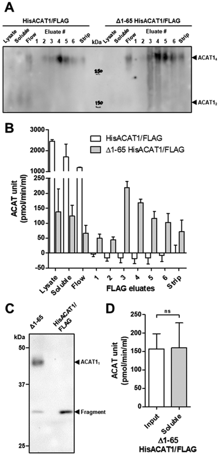
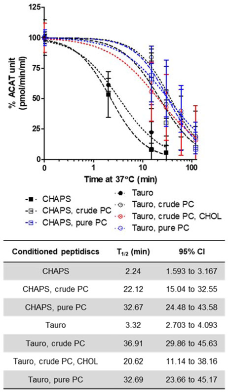
To conduct biochemical studies in vitro, membrane proteins (MPs) must be solubilized with detergents. While detergents are great tools, they can also inhibit the biological activity and/or perturb oligomerization of individual MPs. Nanodisc scaffold peptide (NSPr), an amphipathic peptide analog of ApoA1, was recently shown to reconstitute detergent solubilized MPs into peptidiscs in vitro. Acyl-coenzyme A:cholesterol acyltransferase 1 (ACAT1), also known as sterol O-acyltransferase 1 (SOAT1), plays a key role in cellular cholesterol storage in various cell types and is a drug target to treat multiple human diseases. ACAT1 contains nine transmembrane domains (TMDs) and primarily forms a homotetramer in vitro and in intact cells; deletion of the N-terminal dimerization domain produces a homodimer with full retention in catalytic activity. ACAT1 is prone to inactivation by numerous detergents. Here we pursued the use of NSPr to overcome the detergent-induced inactivation of ACAT1 by generating near detergent-free ACAT1 peptidiscs. Based on native-PAGE analysis, we showed that NSPr reconstitutes ACAT1 into soluble peptidiscs, in which ACAT1 exists predominantly in oligomeric states greater than a homotetramer. The formation of these higher-order oligomeric states was independent of the N-terminal dimerization domain, suggesting that the oligomerization is mediated through hydrophobic interactions of multiple ACAT1 subunits. ACAT1 peptidiscs were still susceptible to heat-mediated inactivation, presumably due to the residual detergent (CHAPS) bound to ACAT1. We then conditioned ACAT1 with phosphatidylcholine (PC) to replace CHAPS prior to the formation of ACAT1 peptidiscs. The results showed, when PC was included, ACAT1 was present mainly in higher-order oligomeric states with greater enzymatic activity. With PC present, the enzymatic activity of ACAT1 peptidiscs was protected from heat-mediated inactivation. These results support the use of NSPr to create a near detergent-free solution of ACAT1 in peptidiscs for various in vitro studies. Our current results also raise the possibility that, under certain conditions, ACAT1 may form higher-order oligomeric states in vivo.

Keywords: ACAT/SOAT; Detergent-free; Membrane protein; Oligomerization; Peptidisc; Phospholipid.


Conflict of interest statement

Conflict of interest

The authors declare that they have no conflicts of interest with the contents of this article.

Figures

Fig. 1
Fig. 1. HisACAT1/FLAG reconstituted into peptidiscs via the in-gel method stabilizes the enzyme in higher-order oligomers.
For this and following figures, Western blots were probed with anti-FLAG-tag antibody for ACAT1 detection. NSPr and pure HisACAT1/FLAG were mixed at the indicated molar ratio before loading onto (A) 10% Clear native (CN) and Blue native (BN)-PAGE or (B) 4-10% gradient CN-PAGE. On-bead HisACAT1/FLAG peptidiscs were eluted from anti-FLAG M2 resin. HisACAT1/FLAG purified to apparent homogeneity was prepared according to [12, 30]. The enzyme preparation was stored at −80°C until usage.
Fig. 2
Fig. 2. HisACAT1/FLAG partially purified in peptidiscs were cleaved and enzymatically inactive, whereas Δ1-65 HisACAT1/FLAG partially purified in peptidiscs were uncleaved and enzymatically active.
This experiment was conducted according to the on-bead reconstitution of ACAT1 peptidiscs method described in section 2.4. (A) Western blot of 4-10% gradient CN-PAGE of HisACAT1/FLAG and Δ1-65 HisACAT1/FLAG peptidiscs. (B) The liposomal ACAT activity of HisACAT1/FLAG and Δ1-65 HisACAT1/FLAG peptidiscs is reported as mean ± standard deviation (SD). (C) Western blot of 10% SDS-PAGE of HisACAT1/FLAG and Δ1-65 HisACAT1/FLAG peptidiscs displaying cleavage of HisACAT1/FLAG to a ~30 kDa fragment. (D) Comparison of liposomal ACAT activity of Δ1-65 HisACAT1/FLAG peptidiscs before (marked as input) and after spinning at 100,000 × g for 30 min at 4°C (marked as soluble) reported as mean ± SD. ns = not significant
Fig. 3
Fig. 3. Purification of Δ1-65 HisACAT1/FLAG with or without NSPr.
The on-bead purification of Δ1-65 HisACAT1/FLAG was conducted as described in section 2.6 and depicted in (A). The ‘X’ denotes the purification of no detectable ACAT1 protein. The composition of various anti-FLAG M2 resin eluants are as indicated. Anti-FLAG-tag antibody was used for all Western blot analyses. (B) Western blot of 4-10% gradient CN-PAGE of Δ1-65 HisACAT1/FLAG with various eluants. (C) Liposomal ACAT activity of Δ1-65 HisACAT1/FLAG with various eluants reported as mean ± SD (n = 3). (D) Western blot of 10% SDS-PAGE of Δ1-65 HisACAT1/FLAG with various eluants. (E) Relative liposomal ACAT activity as compared to Western signal from (D) reported as mean ± SD (n = 3). ns = not significant; * p = ≤ 0.05; ** p = ≤ 0.01; *** p = ≤ 0.0001
Fig. 4
Fig. 4. Conditioning of Δ1-65 HisACAT1/FLAG with CHAPS, or digitonin, or phosphatidylcholine prior to peptidisc reconstitution.
Procedure for on-bead conditioning and reconstitution were described in section 2.7. The conditioning buffers are indicated at the top of the gels. (A) Western blot of 4-10% gradient CN-PAGE loaded with the column eluates. The eluates contained Δ1-65 HisACAT1/FLAG in CHAPS (0.5 M KCl; 8.1 mM CHAPS), or in PC particles (9.3 mM taurocholate; 11.2 mM crude egg PC), or in 0.1 M KCl with exposure to 1 mg/ml NSPr were as indicated. Western blot of (B) 4-10% gradient CN-PAGE or (C) 10% SDS-PAGE loaded with the on-bead reconstitution of Δ1-65 HisACAT1/FLAG peptidiscs in CHAPS, or digitonin (0.5 M KCl; 16.3 μM digitonin), or PC particles prior to the addition of NSPr (E = eluate, S = strip).
Fig. 5
Fig. 5. Conditioning of Δ1-65 HisACAT1/FLAG with detergent micelles (CHAPS or taurocholate), or with several formulations of phosphatidylcholine particles, prior to peptidisc reconstitution.
The composition of conditioning buffers are as follows: CHAPS (0.5 M KCl, 8.1 mM CHAPS); CHAPS, crude PC (9.3 mM CHAPS; 11.2 mM crude egg PC); CHAPS, pure PC (9.3 mM CHAPS; 11.2 mM pure egg PC); Tauro (9.3 mM taurocholate); Tauro, crude PC (9.3 mM taurocholate; 11.2 mM crude egg PC); Tauro, crude PC, CHOL (9.3 mM taurocholate; 11.2 mM crude egg PC; 1.8 mM cholesterol); or Tauro, pure PC (9.3 mM taurocholate; 11.2 mM pure egg PC). (A) Diagram of the experiment where the ‘X’ denotes no protection from heat-mediated inactivation and the ‘check mark’ denotes protection from heat-mediated inactivation. Representative Western blots of (B) 4-10% CN-PAGE or (C) 10% SDS-PAGE of differentially conditioned Δ1-65 HisACAT1/FLAG peptidiscs (1× = 2.5 μl of sample, 2× = 5 μl of sample). (D) Comparison of relative ACAT units to Western blot signal of ACAT1 peptidiscs conditioned with various reagents as indicated (n = 3). Reported as mean ± SD. ** p = ≤ 0.01; *** p = ≤ 0.0001
Fig. 6
Fig. 6. Comparison of the heat-mediated enzymatic inactivation of Δ1-65 HisACAT1/FLAG when present in detergent-conditioned or in phosphatidylcholine-conditioned peptidiscs.
The procedure for conducting heat-mediated inactivation of ACAT1 at 37°C was described in section 2.8. Time course of heat-mediated inactivation of Δ1-65 HisACAT1/FLAG peptidiscs conditioned with CHAPS, or taurocholate, or PC particles (n = 3). Non-linear regression models are reported as log(inhibitor) vs. normalized response with variable slope as determined by GraphPad Prism 5. The half-time of ACAT enzymatic activity decay (T1/2) of the differentially conditioned Δ1-65 HisACAT1/FLAG peptidiscs at 37°C are reported as IC50 ± 95% confidence interval (CI). (n = 3).

Similar articles

Cited by

References

    1. Garavito RM, Ferguson-Miller S, Detergents as tools in membrane biochemistry, The Journal of biological chemistry 276(35) (2001) 32403–6. - PubMed
    1. Bowie JU, Stabilizing membrane proteins, Current opinion in structural biology 11(4) (2001) 397–402. - PubMed
    1. Chang TY, Li BL, Chang CC, Urano Y, Acyl-coenzyme A:cholesterol acyltransferases, American journal of physiology. Endocrinology and metabolism 297(1) (2009) E1–9. - PMC - PubMed
    1. Murphy SR, Chang CC, Dogbevia G, Bryleva EY, Bowen Z, Hasan MT, Chang TY, Acat1 knockdown gene therapy decreases amyloid-beta in a mouse model of Alzheimer's disease, Molecular therapy : the journal of the American Society of Gene Therapy 21(8) (2013) 1497–506. - PMC - PubMed
    1. Yue S, Li J, Lee SY, Lee HJ, Shao T, Song B, Cheng L, Masterson TA, Liu X, Ratliff TL, Cheng JX, Cholesteryl ester accumulation induced by PTEN loss and PI3K/AKT activation underlies human prostate cancer aggressiveness, Cell metabolism 19(3) (2014) 393–406. - PMC - PubMed

Publication types

LinkOut - more resources