. 2020 Oct 15;395(2):112204.
doi: 10.1016/j.yexcr.2020.112204. Epub 2020 Jul 28.

Network perturbation analysis in human bronchial epithelial cells following SARS-CoV2 infection

Affiliations

Network perturbation analysis in human bronchial epithelial cells following SARS-CoV2 infection

Giuseppe Nunnari et al. Exp Cell Res. .

Abstract

Background: SARS-CoV2, the agent responsible for the current pandemic, is also causing respiratory distress syndrome (RDS), hyperinflammation and high mortality. It is critical to dissect the pathogenetic mechanisms in order to reach a targeted therapeutic approach.

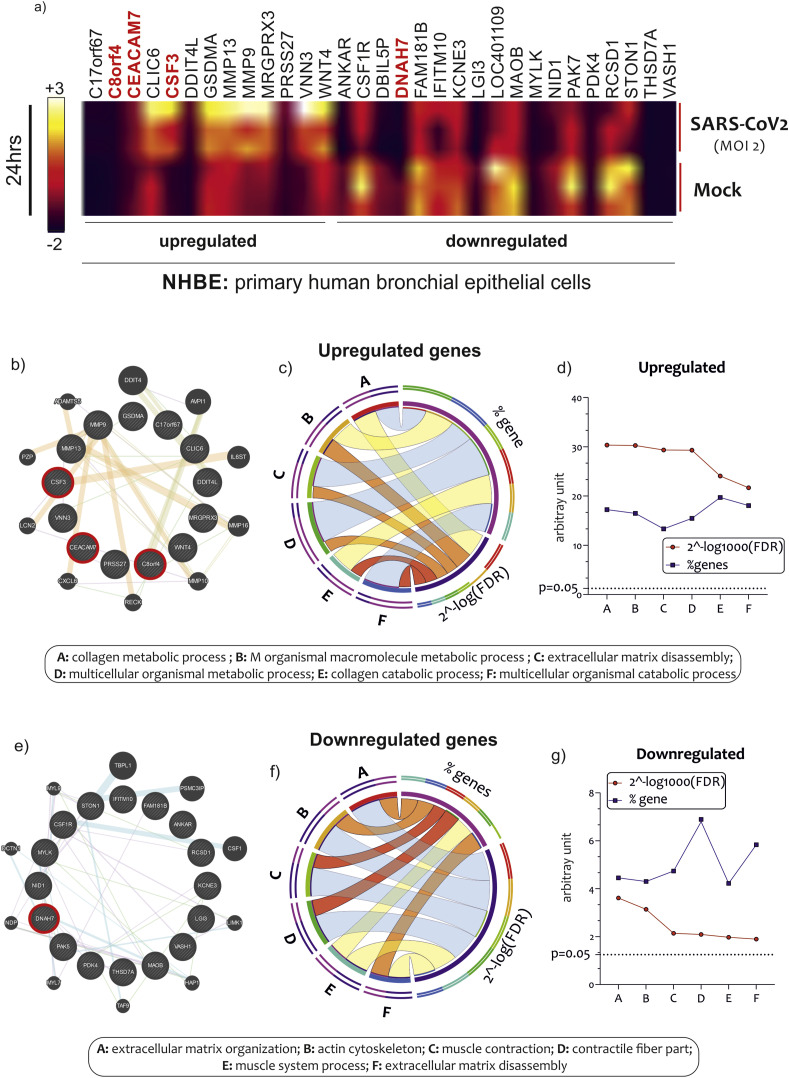
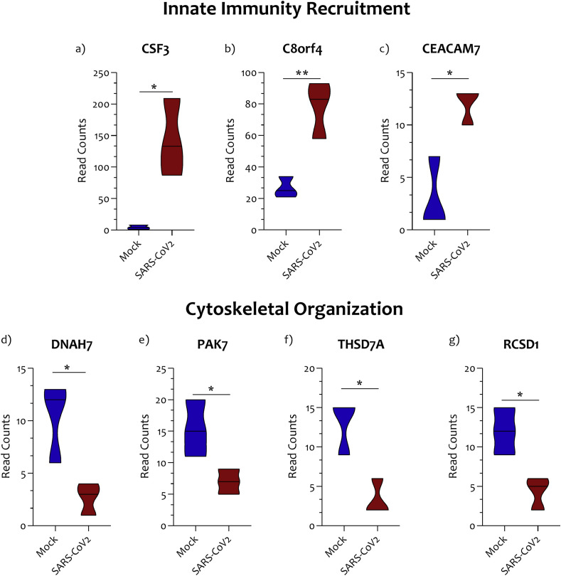
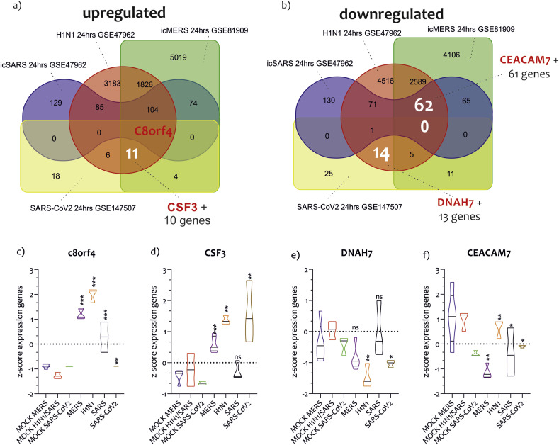
Methods: In the present investigation, we evaluated the effects of SARS-CoV2 on human bronchial epithelial cells (HBEC). We used RNA-seq datasets available online for identifying SARS-CoV2 potential genes target on human bronchial epithelial cells. RNA expression levels and potential cellular gene pathways have been analyzed. In order to identify possible common strategies among the main pandemic viruses, such as SARS-CoV2, SARS-CoV1, MERS-CoV, and H1N1, we carried out a hypergeometric test of the main genes transcribed in the cells of the respiratory tract exposed to these viruses.

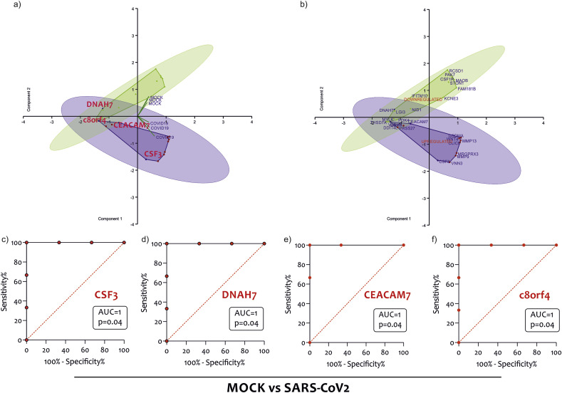
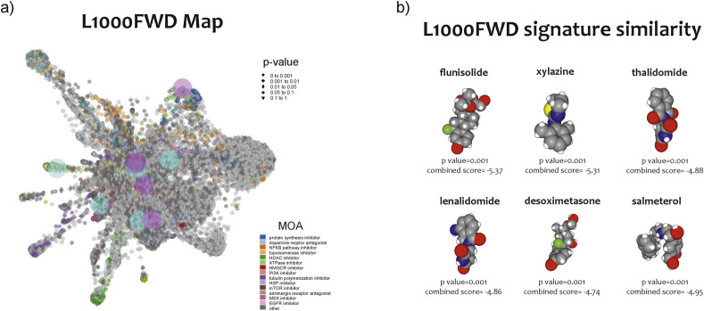
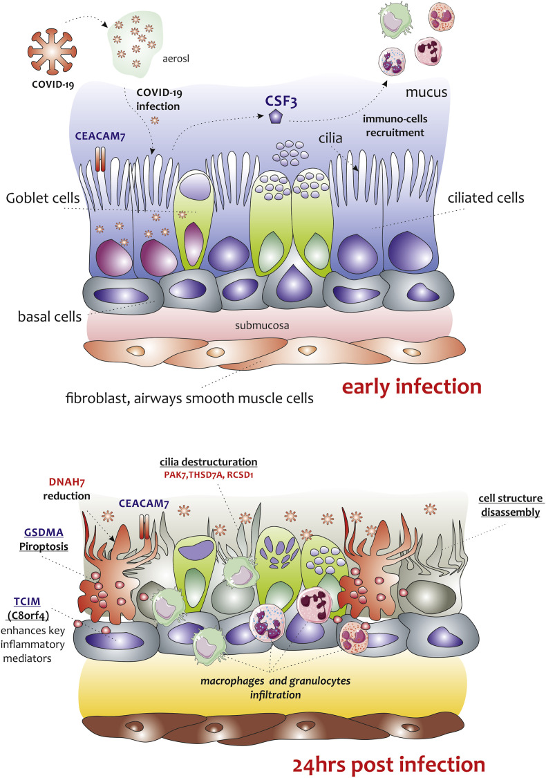
Results: The analysis showed that two mechanisms are highly regulated in HBEC: the innate immunity recruitment and the disassembly of cilia and cytoskeletal structure. The granulocyte colony-stimulating factor (CSF3) and dynein heavy chain 7, axonemal (DNAH7) represented respectively the most upregulated and downregulated genes belonging to the two mechanisms highlighted above. Furthermore, the carcinoembryonic antigen-related cell adhesion molecule 7 (CEACAM7) that codifies for a surface protein is highly specific of SARS-CoV2 and not for SARS-CoV1, MERS-CoV, and H1N1, suggesting a potential role in viral entry. In order to identify potential new drugs, using a machine learning approach, we highlighted Flunisolide, Thalidomide, Lenalidomide, Desoximetasone, xylazine, and salmeterol as potential drugs against SARS-CoV2 infection.

Conclusions: Overall, lung involvement and RDS could be generated by the activation and down regulation of diverse gene pathway involving respiratory cilia and muscle contraction, apoptotic phenomena, matrix destructuration, collagen deposition, neutrophil and macrophages recruitment.

Keywords: Bioinformatics; CEACAM7; COVID-19; CSF3; DNAH7; Innate immunity; Respiratory cilia; SARS-CoV(2); c8orf4.


Conflict of interest statement

The authors declare that they have no known competing financial interests or personal relationships that could have appeared to influence the work reported in this paper.

Figures

Fig. 1
Fig. 1
GSEA of NHBE infected by SARS-CoV2. Heatmap of most upregulated and downregulated genes in NHBE infected with SARS-CoV2 MOI 2, for 24 h. Highlighted in red bold the genes c8orf4, CEACAM7, CSF3, and DNAH7, highly and significantly modulated by the invention (a). GO analysis of 13 and 17 genes statistically significantly upregulated (b) and downregulated (c) in NHBE cells infected with SARS-CoV2. Among these genes, we have excluded for future analysis the c17orf67, ANKAR, LOC401109, and DBIL5P genes because they do not yet have characterized functions. Data are expressed as a string network (GeneMania), Circos graph and Line chart.
Fig. 2
Fig. 2
Innate immunity recruitment and Cytoskeleton-related genes modulated in NHBE cells infected with SARS-CoV2. Three genes out of 12 belong to the “innate immunity recruitment” group, namely the CSF3(a), the transcriptional and immune response regulator (c8orf4) (b), and the carcinoembryonic antigen-related cell adhesion molecule 7 (c). Four gene out of 15 belong to the “mechanisms of cytoskeletal organization”, and were Dynein heavy chain 7 (DNAH7) (d), the P21 (RAC1) activated kinase 5 (PAK7) (e), the thrombospondin type-1 domain-containing protein 7A (THSD7A) (f), and the RCSD domain containing 1 (RCSD1) genes (g). Data are expressed as RNA Read count and presented as violin plots. P values < 0.05 were considered to be statistically significant (*p < 0.05).
Fig. 3
Fig. 3
Gene segregation according to the NHBE cells treatment. Scatterplot of Principal Component Analysis (PCA) using the DEGs highlighted in NHBE treated with COVID-19 from the GSE147507 dataset with (a) and without (b) genes depicted. ROC curve of CSF3 (c), DNAH7 (d), CEACAM7 (e), and c8orf4 (f).
Fig. 4
Fig. 4
Gene signature similarity between SARS-CoV2, SARS-CoV, MERS-CoV and H1N1. Overlap of upregulated genes by SARS-CoV2, SARS-CoV, MERS-CoV, and H1N1 in NHBE and HAE. The analysis showed that c8orf4 was the gene commonly regulated in NHBE and HAE under the infection of the four viruses (a and c). Eleven genes, including CSF3, were commonly modulated by SARS-CoV2, MERS-CoV, and H1N1 (a and d). As regards the overlap of downregulated genes, we showed that no genes were modulated by the four viruses. Fourteen genes were shared between SARS-CoV2 and H1N1, including DNAH7(b and e). 62 genes were identified in common between H1N1, MERS-CoV, and SARS-CoV, including CEACAM7(b and f). Data are expressed as RNA Read count and presented as violin plots. P values < 0.05 were considered to be statistically significant (*p < 0.05).
Fig. 5
Fig. 5
Drugs prediction against SARS-CoV2 infection. L1000FDW visualization of drug-induced signature. Input genes are represented by the significantly upregulated and downregulated genes obtained from the analysis of the GSE147507 dataset. Blue and red circles identify drugs with similar and anti-similar signatures. Dots are color-coded based on the Mode of Action (MOA) of the respective drug (a). The drugs with a high significance pvalue (qvalue) and a high combined score (flunisolide, xylazine, thalidomide, lenalidomide, desoximetasone, and salmeterol) were selected (b).
Fig. 6
Fig. 6
Graphical representation of the main process predicted in NHBE cells infected with SARS-CoV2. Panel A and B show our hypothesis of key events in SARS-CoV2 pathogenesis, which is based on extremely limited observations on an in vitro model of NHBE cell infected with SARS-CoV2 at MOI 2 for 24 h. After the inoculation of SARS-CoV2, the NHBE cells were infected. We hypothesize that viral entry could be facilitated by another cell-surface protein, carcinoembryonic antigen-related cell-adhesion molecule 7 (CEACAM7), which is also expressed in gastrointestinal tract. This mechanism could be similar to that used by the MERS-CoV virus. In that case, the virus uses the CEACAM5 protein to infect the cells of the bronchial epithelium. Inflammatory signaling molecules that are released by infected cells (CSF3, c8orf4), recruits.

References

    1. Bogoch, Watts A., Thomas-Bachli A., Huber C., Kraemer M.U.G., Khan K. Pneumonia of unknown aetiology in Wuhan, China: potential for international spread via commercial air travel. J. Trav. Med. 2020;27 - PMC - PubMed
    1. Lu T., Pu H. Computed tomography manifestations of 5 cases of the novel coronavirus disease 2019 (COVID-19) pneumonia from patients outside Wuhan. J. Thorac. Imag. 2020;35(3):90–93. doi: 10.1097/RTI.0000000000000508. - DOI - PMC - PubMed
    1. Lagunas-Rangel F.A. Neutrophil-to-Lymphocyte ratio and Lymphocyte-to-C-reactive protein ratio in patients with severe coronavirus disease 2019 (COVID-19): a meta-analysis. J. Med. Virol. 2020 doi: 10.1002/jmv.25819. In preparation. - DOI - PMC - PubMed
    1. To K.F., Lo A.W. Exploring the pathogenesis of severe acute respiratory syndrome (SARS): the tissue distribution of the coronavirus (SARS-CoV) and its putative receptor, angiotensin-converting enzyme 2 (ACE2) J. Pathol. 2004;203:740–743. - PMC - PubMed
    1. Clough E., Barrett T. The gene expression Omnibus database. Methods Mol. Biol. 2016;1418:93–110. - PMC - PubMed

MeSH terms