Review
. 2021 Mar:81:100883.
doi: 10.1016/j.preteyeres.2020.100883. Epub 2020 Jul 28.

TCF4-mediated Fuchs endothelial corneal dystrophy: Insights into a common trinucleotide repeat-associated disease

Affiliations
Review

TCF4-mediated Fuchs endothelial corneal dystrophy: Insights into a common trinucleotide repeat-associated disease

Michael P Fautsch et al. Prog Retin Eye Res. 2021 Mar.

Abstract

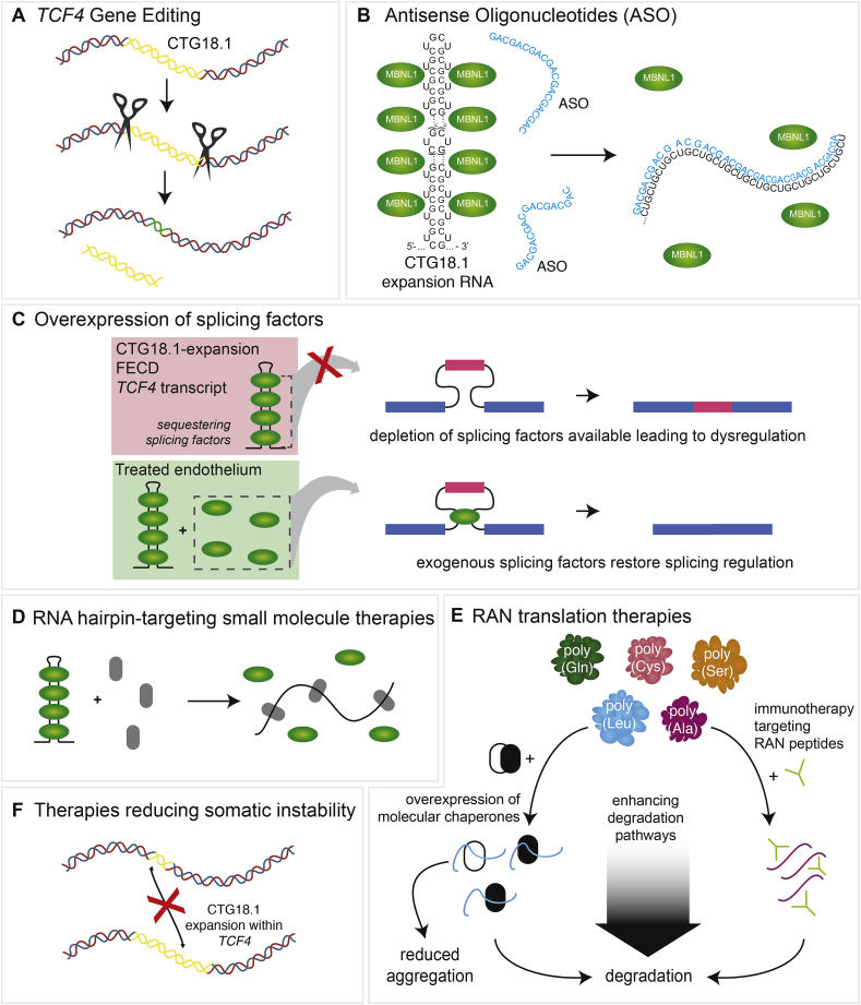
Fuchs endothelial corneal dystrophy (FECD) is a common cause for heritable visual loss in the elderly. Since the first description of an association between FECD and common polymorphisms situated within the transcription factor 4 (TCF4) gene, genetic and molecular studies have implicated an intronic CTG trinucleotide repeat (CTG18.1) expansion as a causal variant in the majority of FECD patients. To date, several non-mutually exclusive mechanisms have been proposed that drive and/or exacerbate the onset of disease. These mechanisms include (i) TCF4 dysregulation; (ii) toxic gain-of-function from TCF4 repeat-containing RNA; (iii) toxic gain-of-function from repeat-associated non-AUG dependent (RAN) translation; and (iv) somatic instability of CTG18.1. However, the relative contribution of these proposed mechanisms in disease pathogenesis is currently unknown. In this review, we summarise research implicating the repeat expansion in disease pathogenesis, define the phenotype-genotype correlations between FECD and CTG18.1 expansion, and provide an update on research tools that are available to study FECD as a trinucleotide repeat expansion disease. Furthermore, ongoing international research efforts to develop novel CTG18.1 expansion-mediated FECD therapeutics are highlighted and we provide a forward-thinking perspective on key unanswered questions that remain in the field.

Keywords: CTG18.1; FECD; Fuchs endothelial corneal dystrophy; RAN translation; RNA toxicity; Repeat-expansion; Transcription factor 4; Trinucleotide repeat; Triplet repeat-mediated disease.


Conflict of interest statement

MPF, EDW, KHB, NB, ANS, NJHT, SJT – no conflicts of interest.

AED is a member of Triplet Therapeutics Scientific Advisory board. Research conducted in AED's laboratory is partly funded by ProQR Therapeutics.

Figures

Fig. 1
Fig. 1
Clinical images of Fuchs endothelial corneal dystrophy (FECD). A. Edema of the central cornea due to FECD. B. Retro-illumination photograph of a cornea with FECD showing numerous guttae that give a stippled appearance. C. Periodic acid-Schiff (PAS)-stained cross-section of normal cornea (endothelial cells, arrow; Descemet's membrane, asterisk). D. PAS-stained cross-section of cornea with FECD showing posterior elevations (guttae, arrows) arising from the thickened Descemet membrane (asterisk). E. In vivo confocal image of normal corneal endothelial cells show a regular hexagonal pattern. F. In vivo confocal image of FECD corneal endothelium with disrupted hexagonal cell pattern. Dark areas represent guttae.
Fig. 2
Fig. 2
Manhattan plot illustrating a genome wide significant association between Fuchsendothelialcornealdystrophy (FECD) and a region on chromosome 18, encompassing TCF4. Genome wide association study (GWAS) comparing 338,727 Single-Nucleotide Polymorphisms (SNPs) and FECD. The negative log of the P values of association between genotyped SNPs and an FECD discovery cohort is plotted against chromosomal location. One region on chromosome 18, encompassing TCF4, was found to reach genome wide significance (p = 1.0 × 10−12) within this discovery cohort. Used with permission from Baratz et al. (2010).
Fig. 3
Fig. 3
Mechanisms of cellular dysregulation associated with CTG18.1 expansions. Four non-mutually exclusive mechanisms have been proposed to drive and/or exacerbate the onset of CTG18.1 expansion-mediated FECD, including; (1) dysregulated expression of TCF4 transcripts, (2) accumulation of toxic (a) sense (CUG)n and (b) antisense-derived (CAG)n repetitive RNA transcripts, (3) RAN translation of repetitive RNA transcripts, and (4) age and tissue-dependant somatic instability of the repeat element.
Fig. 4
Fig. 4
Schematic illustration of TCF4 genomic sequence encompassing CTG18.1, defined protein coding TCF4 isoforms, and characterized TCF4 functional domains. A. Genomic sequence surrounding the CTG18.1 repeat element (red) and adjacent CTC repeat (blue). Intronic sequence surrounding the repeat is shown in lowercase black lettering. Exonic sequence is represented by uppercase letters. B. A schematic of the canonical TCF4 transcript (ENST00000354452.8; NM_001083962.2) encoding Isoform B (ENSP00000346440.3). Arrowhead shows the relative position of the SNP rs613872. Sequence is presented in a 5′ to 3′ orientation. C. A schematic representation of all characterized, protein coding, TCF4 transcripts reported in Ensembl. Transcripts that contain or are in close proximity to the CTG18.1 repeat (red dotted box) are grouped and colored in black. Those transcripts which do not encompass the repeat are colored grey. The transcripts encoding isoform A (TCF4-204 – red) and Isoform B (TCF4-201 - blue) are labelled. Colored boxes denote exons which code for specific TCF4 functional domains. D. Schematic of TCF4 protein functional domains characterized within isoform A and isoform B. Activation domain 1 (AD1) and activation domain 2 (AD2) are represented in blue, whereas the nuclear localization signal (NLS) is in red. Repressor regions of the protein are represented with R. The basic helix-loop-helix (bHLH) domain and the C domain are highlighted in light green and dark green, respectively.
Fig. 5
Fig. 5
Representative fluorescence in situ hybridization (FISH) images of healthy control, CTG18.1 expansion-negative Fuchs endothelial corneal dystrophy (FECD) and CTG18.1 expansion-positive FECD corneal explant tissue. (CUG)n RNA foci detection was performed using Cy3-(CAG)7 probe using methods adapted from Zarouchlioti et al., (2018). White arrows highlight RNA foci within the CTG18.1 expansion-positive endothelium. y.o., years old. Scale bar for multiple nuclei, 20 μm (top panel). Single nuclei, 5 μm (lower panels).
Fig. 6
Fig. 6
Sequestration of MBNL1 to RNA foci within corneal explant tissue. A. MBNL1 is sequestered to the RNA foci within the corneal endothelium of only CTG18.1 expansion-positive FECD patient-derived tissues, scale bar 5 μM. B. Schematic illustrating that in the absence of CTG18.1 expansions, MBNL1 is soluble within the nucleus. C. In the presence of a CTG18.1 expansion, levels of functional MBNL1 are depleted due to the sequestration of the splicing factor to hairpin structures formed by the RNAs comprising expanded copies of TCF4 transcripts. D. Schematic depicting hypothesised mechanism of splicing dysregulation observed within expansion-positive corneal endothelial cells where the sequestering of splicing factors to hairpin structures formed by TCF4 RNA transcripts results in widespread dysregulation of pre-mRNA splicing. Permission for re-use of (A) from Du et al. (2015) was granted through CC-BY license (https://creativecommons.org/licenses/by/4.0/legalcode).
Fig. 7
Fig. 7
Poly-peptide proteins potentially translated from CTG18.1 expansions. Illustration of the potential repeat peptides that may be generated by repeat-associated non-AUG (RAN) translation from expanded (CUG)n RNA transcripts. The sense strand sequence may generate poly-Leucine (Leu), poly-Alanine (Ala) and poly-Cysteine (Cys) peptides and the antisense strand sequence may generate poly-Glutamine (Gln), poly-Alanine (Ala) and poly-Serine (Ser) peptides.
Fig. 8
Fig. 8
Primary corneal endothelial cell (CEC) cultures provide an in vitro system to model CTG18.1 expansion-associated Fuchs endothelial corneal dystrophy (FECD) and display key biomarkers synonymous with CTG18.1-expansion associated FECD within corneal explant tissue. A. Schematic depicting the approach to culture primary human CECs from corneal explant tissue. B. Phase contrast image of primary CECs in culture displaying hexagonal morphology. Scale bar, 0.4 mm. C. Primary CEC cultures displaying distinctive corneal endothelial polygonal morphology and expressing markers indicative of endothelial cell status. Endothelial markers N-Cadherin, ZO-1, ATP1A1, N-CAM and CD166 were detected and the epithelial marker E-Cadherin was absent in CEC lines derived from healthy explant tissue. Scale bars, 100 μm. D. Representative images of MBNL1 protein nuclear localization in CEC derived from CTG18.1 expansion-positive FECD-affected subjects and CTG18.1 expansion-negative FECD-affected subjects. RNA foci are labelled with a Cy3-(CAG)7 probe and DAPI is used to stain nuclei. Scale bars, 10 μm. E. Aberrantly regulated pre-mRNA splicing events are detected within CTG18.1 expansion-positive primary CECs. Graphs presented represent the mean percentage expression of amplicons of interest relative to total amplified products, per reaction, for each respective group for MBNL1, MBNL2, and NUMA1 transcripts. Error bars represent ±1 standard deviation. P values were calculated by one-way analysis of variance (ANOVA); ∗P < 0.001. Permission for re-use of adapted figure from Zarouchlioti et al., (2018) was granted through CC-BY license (https://creativecommons.org/licenses/by/4.0/legalcode).
Fig. 9
Fig. 9
Potential therapeutic strategies targeting pathogenic mechanisms associated with CTG18.1 expansion-mediated Fuchs endothelial corneal dystrophy (FECD). A. Gene editing tools may be applied in the future to reduce the size or presence of CTG18.1 expansions. The CTG18.1 repeat element is represented as yellow. B. Antisense oligonucleotide (ASO) therapeutic strategies can target (CUG)n or (CAG)n RNA transcripts derived from expanded copies of CTG18.1 to physically block the formation of foci and or induce degradation of such transcripts. C. Overexpression of splicing factors, such as MBNL proteins, could restore splicing regulation. D. Small molecule therapeutics (grey) could induce disruption of RNA hairpins on (CUG)n or (CAG)n RNA transcripts, releasing sequestered splicing factors (green). E. Various strategies targeting RAN translation may in the future prove to have therapeutic benefit. Immunotherapy targeting RAN peptides would enable the cell to degrade the peptide aggregates. Overexpression of molecular chaperones could potentially prevent the aggregation of RAN peptides and/or enhance degradation levels. F. Therapeutics aimed at reducing levels of somatic and age-related levels of repeat instability may in the future prove to have therapeutic benefit for CTG18.1-expansion associated FECD.

References

    1. Afshari N.A., Igo R.P., Jr., Morris N.J., Stambolian D., Sharma S., Pulagam V.L., Dunn S., Stamler J.F., Truitt B.J., Rimmler J., Kuot A., Croasdale C.R., Qin X., Burdon K.P., Riazuddin S.A., Mills R., Klebe S., Minear M.A., Zhao J., Balajonda E., Rosenwasser G.O., Baratz K.H., Mootha V.V., Patel S.V., Gregory S.G., Bailey-Wilson J.E., Price M.O., Price F.W., Jr., Craig J.E., Fingert J.H., Gottsch J.D., Aldave A.J., Klintworth G.K., Lass J.H., Li Y.J., Iyengar S.K. Genome-wide association study identifies three novel loci in Fuchs endothelial corneal dystrophy. Nat. Commun. 2017;8:14898. - PMC - PubMed
    1. Afshari N.A., Pittard A.B., Siddiqui A., Klintworth G.K. Clinical study of Fuchs corneal endothelial dystrophy leading to penetrating keratoplasty: a 30-year experience. Arch. Ophthalmol. 2006;124:777–780. - PubMed
    1. Amiel J., Rio M., de Pontual L., Redon R., Malan V., Boddaert N., Plouin P., Carter N.P., Lyonnet S., Munnich A., Colleaux L. Mutations in TCF4, encoding a class I basic helix-loop-helix transcription factor, are responsible for Pitt-Hopkins syndrome, a severe epileptic encephalopathy associated with autonomic dysfunction. Am. J. Hum. Genet. 2007;80:988–993. - PMC - PubMed
    1. Angelbello A.J., DeFeo M.E., Glinkerman C.M., Boger D.L., Disney M.D. Precise targeted cleavage of a r(CUG) repeat expansion in cells by using a small-molecule-deglycobleomycin conjugate. ACS Chem. Biol. 2020;15:849–855. - PMC - PubMed
    1. Angelbello A.J., Rzuczek S.G., McKee K.K., Chen J.L., Olafson H., Cameron M.D., Moss W.N., Wang E.T., Disney M.D. Precise small-molecule cleavage of an r(CUG) repeat expansion in a myotonic dystrophy mouse model. Proc. Natl. Acad. Sci. U. S. A. 2019;116:7799–7804. - PMC - PubMed

Publication types

MeSH terms