. 2020 Jul 24;7(5):e817.
doi: 10.1212/NXI.0000000000000817. Print 2020 Sep.

Antiparanodal antibodies and IgG subclasses in acute autoimmune neuropathy

Affiliations

Antiparanodal antibodies and IgG subclasses in acute autoimmune neuropathy

Luise Appeltshauser et al. Neurol Neuroimmunol Neuroinflamm. .

Abstract

Objective: To determine whether IgG subclasses of antiparanodal autoantibodies are related to disease course and treatment response in acute- to subacute-onset neuropathies, we retrospectively screened 161 baseline serum/CSF samples and 66 follow-up serum/CSF samples.

Methods: We used ELISA and immunofluorescence assays to detect antiparanodal IgG and their subclasses and titers in serum/CSF of patients with Guillain-Barré syndrome (GBS), recurrent GBS (R-GBS), Miller-Fisher syndrome, and acute- to subacute-onset chronic inflammatory demyelinating polyradiculoneuropathy (A-CIDP). We evaluated clinical data retrospectively.

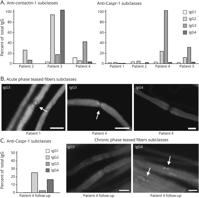
Results: We detected antiparanodal autoantibodies with a prevalence of 4.3% (7/161), more often in A-CIDP (4/23, 17.4%) compared with GBS (3/114, 2.6%). Longitudinal subclass analysis in the patients with GBS revealed IgG2/3 autoantibodies against Caspr-1 and against anti-contactin-1/Caspr-1, which disappeared at remission. At disease onset, patients with A-CIDP had IgG2/3 anti-Caspr-1 and anti-contactin-1/Caspr-1 or IgG4 anti-contactin-1 antibodies, IgG3 being associated with good response to IV immunoglobulins (IVIg). In the chronic phase of disease, IgG subclass of one patient with A-CIDP switched from IgG3 to IgG4.

Conclusion: Our data (1) confirm and extend previous observations that antiparanodal IgG2/3 but not IgG4 antibodies can occur in acute-onset neuropathies manifesting as monophasic GBS, (2) suggest association of IgG3 to a favorable response to IVIg, and (3) lend support to the hypothesis that in some patients, an IgG subclass switch from IgG3 to IgG4 may be the correlate of a secondary progressive or relapsing course following a GBS-like onset.


Figures

Figure 1
Figure 1. Detection of antiparanodal autoantibodies by binding assays on teased fibers, CBA, and preabsorption assay
(A) Colocalization overlay photomicrographs of double immunofluorescence assays with commercial autoantibody displayed in red and human serum in green. Colocalization at the paranodes (arrows) appears yellow and indicates antiparanodal autoantibodies in patients 1–5 (A.b–A.f), but not in the negative control serum (A.a). (B) The specific target of the autoantibodies is illustrated by colocalization overlay photomicrographs on transfected HEK293 cells (arrowheads) with commercial antibody in red, serum in green, and nucleus staining in blue. Colocalization appears yellow (merge) and confirms anti–contactin-1 in patients 2 (B.c), 3 (B.d), and 4 (B.e), but not in patients 1 (B.b), 5 (B.f), and the negative control (B.a). Sera of patients 1, 2, 4, and 5 (B.h, B.i, B.k, and B.l) colocalize on Caspr-1 (CNTNAP1)–transfected HEK293 cells as proof of anti–Caspr-1 autoantibodies, whereas sera of the negative control (B.g) and patient 3 (B.j) do not show anti–Caspr-1 positivity. (C) Single immunofluorescence on murine teased fibers after HEK293 contactin-1 and Caspr-1 preabsorption shows that antiparanodal autoantibodies disappear after contactin-1 preabsorption in patient 3 (C.d) and after Caspr-1 preabsorption in patient 4 (C.f), proving specificity to the respective target antigen. Scale bar = 10 µm. CBA = cell-based assay; HEK = human embryonic kidney.
Figure 2
Figure 2. Serum subclass analysis at baseline and at follow-up with IgG subclass switch
(A) Results of anti–contactin-1 and anti–Caspr-1 ELISA with baseline sera and subclass-specific IgG1-4 autoantibodies (grayscale) are shown as % of the OD of the total IgG (using anti-human IgG autoantibody) on y-axis. In patient 1, ELISA IgG subclasses were not measurable. ELISA revealed mainly IgG2 subclass in patient 2 (GBS), IgG2/IgG4 in patient 3 (A-CIDP), and IgG2/3 in patients 4 and 5 (A-CIDP). (B) Acute phase teased fibers subclass analysis with baseline sera showed binding to the paranodal regions (arrows) only when using IgG3-specific secondary antibody in patients 1 (GBS) and 4 (A-CIDP), visualized by photomicrographs of single immunofluorescence staining on teased fibers. (C) Anti–Caspr-1 IgG subclass ELISA with follow-up serum of patient 4 now revealed IgG 2/4 subclass. Photomicrographs show immunofluorescence staining of murine teased fibers with subclass-specific IgG3 and IgG4 autoantibodies at follow-up. Paranodal binding disappeared using IgG3 antibody, but occurred using IgG4 subclass-specific antibody, proving subclass switch in patient 4. Scale bar = 10 µm. A-CIDP = acute-onset chronic inflammatory demyelinating polyradiculoneuropathy; GBS = Guillain-Barré syndrome.
Figure 3
Figure 3. Antiparanodal IgG autoantibodies at follow-up assessment
(A) Contactin-1 IgG ELISA of all patients at first assessment, of 66/161 patients at follow-up, and of 40 healthy controls. Optical density (OD) at 450 nm is displayed on the y-axis. The threshold for positive results is set at 5 SDs above the mean of controls (0.615). Patients 2–4 show positive results at first assessment. The other patients and controls show values below the threshold. Sera of patients 2, 4, and 5 are negative in anti–contactin-1 IgG ELISA at follow-up, as well as 63 other follow-up sera. Patients 1 and 3 were lost to follow-up. (B) Overlay photomicrographs of teased fibers double immunofluorescence and (C) contactin-1– and Caspr-1 (CNTNAP1)–transfected HEK293 cells show loss of anti–contactin-1/Caspr-1 autoantibodies in patient 2 (GBS) at follow-up (B.a, C.a, C.d), but colocalization of commercial anti–Caspr-1 antibody and sera at paranodes in patients 4 and 5 with A-CIDP (B.b, B.c, arrows). Both sera bind to Caspr-1 (CNTNAP1)–transfected HEK293 cells (C.e, C.f, arrowheads), but not to contactin-1–transfected cells (C.b, C.c). Scale bar = 10 µm. A-CIDP = acute-onset chronic inflammatory demyelinating polyradiculoneuropathy; GBS = Guillain-Barré syndrome; HEK = human embryonic kidney.
Figure 4
Figure 4. Scheme of the autoantibody status in the course of disease in seropositive patients
The course of disease is shown on the x-axis by pseudo-logarithmic display of the time course. Time point 0 is set at baseline assessment of serum/CSF. The color code indicates severity of symptoms. Results of serum assessment are displayed in black. Patients 1 and 3 were lost to follow-up.

Comment in

  • Neurol Neuroimmunol Neuroinflamm. 7(5):e843.

References

    1. Fehmi J, Scherer SS, Willison HJ, Rinaldi S. Nodes, paranodes and neuropathies. J Neurol Neurosurg Psychiatry 2018;89:61–71. - PubMed
    1. Uncini A, Vallat JM. Autoimmune nodo-paranodopathies of peripheral nerve: the concept is gaining ground. J Neurol Neurosurg Psychiatry 2018;89:627–635. - PubMed
    1. Querol L, Illa I. Paranodal and other autoantibodies in chronic inflammatory neuropathies. Curr Opin Neurol 2015;28:474–479. - PubMed
    1. Querol L, Rojas-Garcia R, Diaz-Manera J, et al. . Rituximab in treatment-resistant CIDP with antibodies against paranodal proteins. Neurol Neuroimmunol Neuroinflamm 2015;2:e149. doi: 10.1212/NXI.0000000000000149. - DOI - PMC - PubMed
    1. Querol L, Nogales-Gadea G, Rojas-Garcia R, et al. . Neurofascin IgG4 antibodies in CIDP associate with disabling tremor and poor response to IVIg. Neurology 2014;82:879–886. - PMC - PubMed

Publication types

MeSH terms