Meta-Analysis
. 2020 Jul 31;17(1):227.
doi: 10.1186/s12974-020-01898-y.

Meta-analysis of mouse transcriptomic studies supports a context-dependent astrocyte reaction in acute CNS injury versus neurodegeneration

Affiliations
Meta-Analysis

Meta-analysis of mouse transcriptomic studies supports a context-dependent astrocyte reaction in acute CNS injury versus neurodegeneration

Sudeshna Das et al. J Neuroinflammation. .

Abstract

Background: Neuronal damage in acute CNS injuries and chronic neurodegenerative diseases is invariably accompanied by an astrocyte reaction in both mice and humans. However, whether and how the nature of the CNS insult-acute versus chronic-influences the astrocyte response, and whether astrocyte transcriptomic changes in these mouse models faithfully recapitulate the astrocyte reaction in human diseases remains to be elucidated. We hypothesized that astrocytes set off different transcriptomic programs in response to acute versus chronic insults, besides a shared "pan-injury" signature common to both types of conditions, and investigated the presence of these mouse astrocyte signatures in transcriptomic studies from human neurodegenerative diseases.

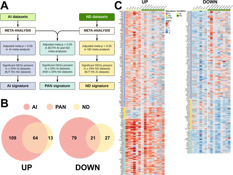
Methods: We performed a meta-analysis of 15 published astrocyte transcriptomic datasets from mouse models of acute injury (n = 6) and chronic neurodegeneration (n = 9) and identified pan-injury, acute, and chronic signatures, with both upregulated (UP) and downregulated (DOWN) genes. Next, we investigated these signatures in 7 transcriptomic datasets from various human neurodegenerative diseases.

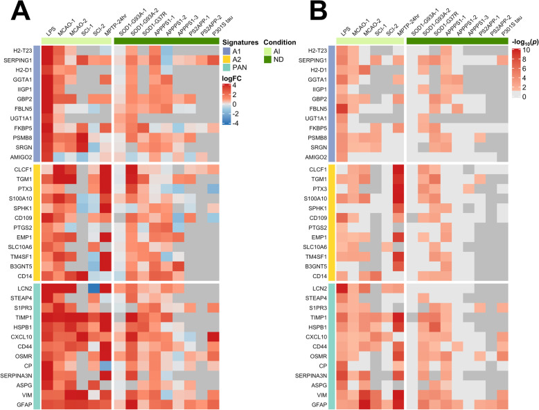
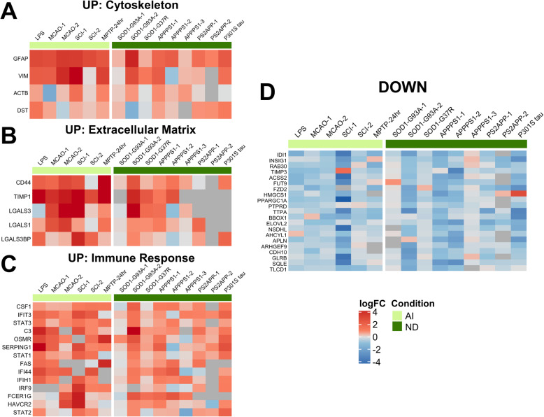
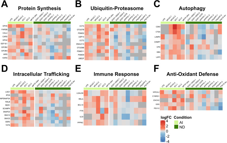
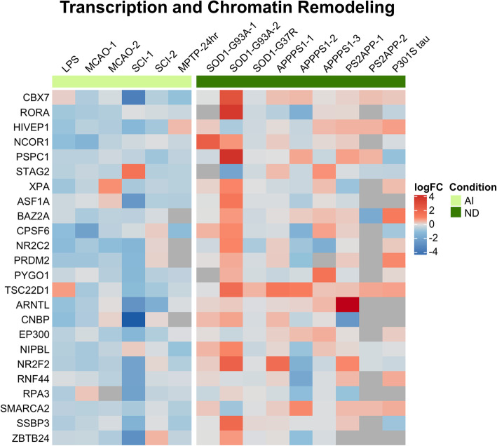
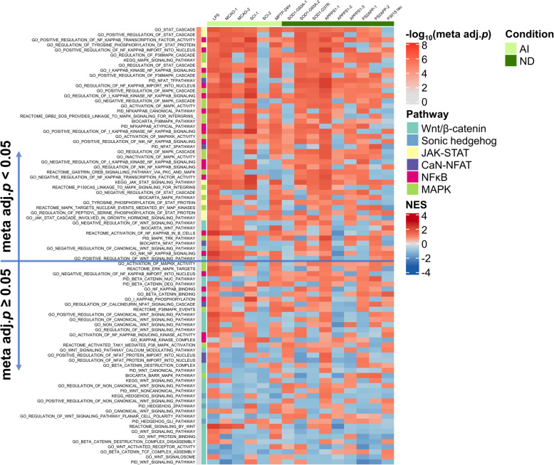
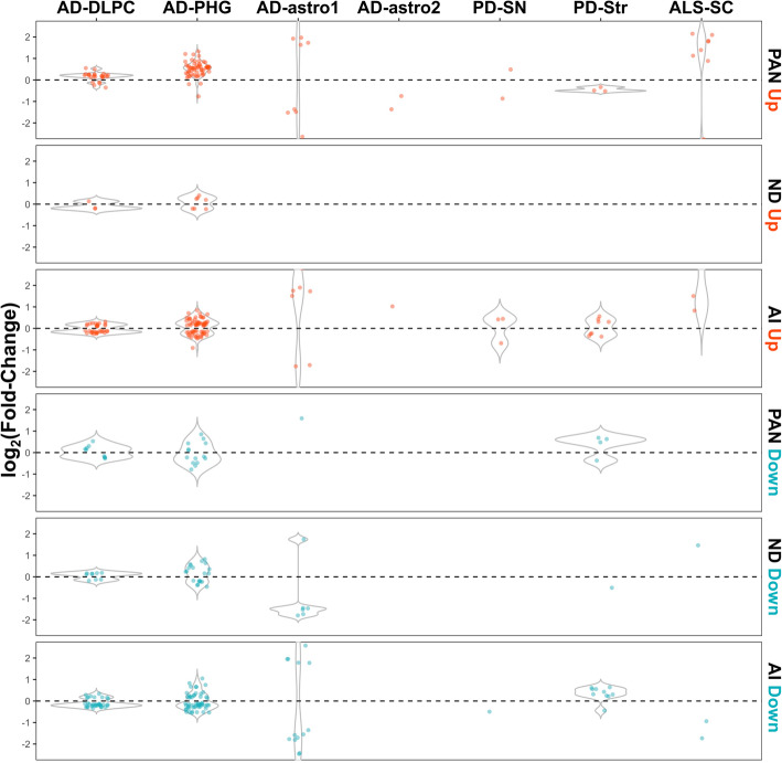
Results: In mouse models, the number of UP/DOWN genes per signature was 64/21 for pan-injury and 109/79 for acute injury, whereas only 13/27 for chronic neurodegeneration. The pan-injury-UP signature was represented by the classic cytoskeletal hallmarks of astrocyte reaction (Gfap and Vim), plus extracellular matrix (i.e., Cd44, Lgals1, Lgals3, Timp1), and immune response (i.e., C3, Serping1, Fas, Stat1, Stat2, Stat3). The acute injury-UP signature was enriched in protein synthesis and degradation (both ubiquitin-proteasome and autophagy systems), intracellular trafficking, and anti-oxidant defense genes, whereas the acute injury-DOWN signature included genes that regulate chromatin structure and transcriptional activity, many of which are transcriptional repressors. The chronic neurodegeneration-UP signature was further enriched in astrocyte-secreted extracellular matrix proteins (Lama4, Cyr61, Thbs4), while the DOWN signature included relevant genes such as Agl (glycogenolysis), S1pr1 (immune modulation), and Sod2 (anti-oxidant). Only the pan-injury-UP mouse signature was clearly present in some human neurodegenerative transcriptomic datasets.

Conclusions: Acute and chronic CNS injuries lead to distinct astrocyte gene expression programs beyond their common astrocyte reaction signature. However, caution should be taken when extrapolating astrocyte transcriptomic findings from mouse models to human diseases.

Keywords: Acute CNS injury; Astrocyte reaction; Meta-analysis; Neurodegenerative diseases; Transcriptomics.


Conflict of interest statement

The authors declare that they have no competing interests.

Figures

Fig. 1
Fig. 1
Purported neurotoxic, neuroprotective, and pan-reactive astrocyte transcriptomic signatures in acute injury and chronic neurodegenerative mouse models. Heatmaps illustrate the log2(fold-change) (a) and − log10(p values) (b) of the neurotoxic (A1), neuroprotective (A2), and pan-reactive (PAN) gene cassettes as defined by Liddelow et al. [2]. Dark gray in (a) and (b) means that the transcript was not detected or was detected at extremely low levels. Light gray transcripts in (b) were not statistically significant. Note that all three signatures are significantly upregulated in astrocytes from chronic neurodegeneration (ND) and, specially, acute injury (AI) mouse models
Fig. 2
Fig. 2
Meta-analysis shows distinct acute injury and chronic neurodegeneration signatures, and a common pan-injury signature. a Flow-chart shows the criteria used to develop acute injury (AI), chronic neurodegenerative diseases (ND), and pan-injury (PAN) signatures. b Venn diagrams show the number of upregulated (UP) and downregulated (DOWN) genes for each of the signatures. c Heatmaps depict log2(fold change) of upregulated (UP) and downregulated (DOWN) genes comprising the three signatures
Fig. 3
Fig. 3
Pan-injury astrocyte gene expression signature. Heatmaps depict log2(fold change) of upregulated (ac) and downregulated (d) genes in both acute injury (AI) and chronic neurodegeneration (ND). The pan-injury (PAN) signature consists of upregulation of cytoskeleton (i.e., Gfap, Vim) (a), extracellular matrix (i.e., Cd44, Timp1) (b), and immune response (i.e., C3, interferon signaling, STAT pathway) (c), and downregulation of lipid metabolism (i.e., Hmgcs1, Sqle) among other genes (d)
Fig. 4
Fig. 4
Astrocyte gene expression signature upregulated in acute injury. Heatmaps depict log2(fold change) of acute injury-specific upregulated genes. The upregulated acute injury signature consists of protein translation (i.e., elongation machinery and aminoacyl tRNA synthetases) (a), ubiquitin-proteasome system (i.e., proteasome subunits and chaperones) (b), autophagy system (i.e., lysosomal enzymes such as cathepsin A and glucocerebrosidase, and lysosomal associated membrane protein 2) (c), intracellular trafficking (i.e., ER-Golgi) (d), immune response (i.e., Rela, Il33) (e), and anti-oxidant defenses (i.e., Nfe2l2, Gss)
Fig. 5
Fig. 5
Astrocyte gene expression signature downregulated in acute injury. Downregulated genes in acute injury were mainly related to chromatin remodeling and organization and transcription, including many transcriptional repressors
Fig. 6
Fig. 6
Astrocyte gene expression signature in chronic neurodegeneration. The chronic neurodegeneration signature is characterized by upregulation (a) of amyloid precursor protein, calpain-3, and secreted matricellular proteins (i.e., Cyr61, Lama4, Thbs4), and downregulation (b) of metabolic processes (i.e., Acot6, Agl, Naa30)
Fig. 7
Fig. 7
Inflammatory but not proliferation signaling pathways are upregulated in acute injury and chronic neurodegeneration. Heatmap shows the normalized enrichment score (NES) of pro-inflammatory (NFκB, calcineurin/NFAT, MAPK, and JAK/STAT) and proliferative (Wnt/β-catenin and sonic hedgehog) gene sets in the astrocyte transcriptomic datasets analyzed. The gene sets are color-coded by pathway and ordered by descending meta-analytic p value [− log10(Meta adj. p)]. Note that NFκB, MAPK, and JAK/STAT gene sets are more significantly upregulated than Wnt/β-catenin and sonic hedgehog
Fig. 8
Fig. 8
Validation of mouse astrocyte transcriptomic signatures in human neurodegenerative transcriptomic datasets. Violin plots represent the logFC of statistically significant DEGs (p < 0.05) from the acute injury (AI), chronic neurodegenerative (ND), and pan-injury (PAN) mouse astrocyte signatures in diseased versus control human subjects. Note that only the two AD bulk tissue datasets (AD-DLPC and AD-PHG) showed some concordance with the pan-injury-UP mouse astrocyte signatures, but not with any of the others. Abbreviations: AD-DLPC, AD dorsolateral prefrontal cortex from ROSMAP; AD-PHG, AD parahippocampal gyrus from MSBB; AD-astro1, GFAP+ astrocytes laser capture microdissected from lateral temporal cortex; AD-astro2, GFAP+ astrocytes sorted from superior frontal cortex; ALS-SC, amyotrophic lateral sclerosis spinal cord gray matter; PD-SN, Parkinson’s disease substantia nigra; PD-Str, Parkinson’s disease striatum

References

    1. Zamanian JL, Xu L, Foo LC, Nouri N, Zhou L, Giffard RG, et al. Genomic analysis of reactive astrogliosis. J Neurosci Off J Soc Neurosci. 2012;32(18):6391–6410. - PMC - PubMed
    1. Liddelow SA, Guttenplan KA, Clarke LE, Bennett FC, Bohlen CJ, Schirmer L, et al. Neurotoxic reactive astrocytes are induced by activated microglia. Nature. 2017;541(7638):481–487. - PMC - PubMed
    1. Buffo A, Rite I, Tripathi P, Lepier A, Colak D, Horn A-P, et al. Origin and progeny of reactive gliosis: a source of multipotent cells in the injured brain. Proc Natl Acad Sci U S A. 2008;105(9):3581–3586. - PMC - PubMed
    1. Bardehle S, Krüger M, Buggenthin F, Schwausch J, Ninkovic J, Clevers H, et al. Live imaging of astrocyte responses to acute injury reveals selective juxtavascular proliferation. Nat Neurosci. 2013;16(5):580–586. - PubMed
    1. Sirko S, Behrendt G, Johansson PA, Tripathi P, Costa M, Bek S, et al. Reactive glia in the injured brain acquire stem cell properties in response to sonic hedgehog. [corrected] Cell Stem Cell. 2013;12(4):426–439. - PubMed

Publication types

Substances