. 2020 Sep 1;205(5):1449-1460.
doi: 10.4049/jimmunol.2000007. Epub 2020 Jul 31.

Adoptive T Cell Therapy with IL-12-Preconditioned Low-Avidity T Cells Prevents Exhaustion and Results in Enhanced T Cell Activation, Enhanced Tumor Clearance, and Decreased Risk for Autoimmunity

Affiliations

Adoptive T Cell Therapy with IL-12-Preconditioned Low-Avidity T Cells Prevents Exhaustion and Results in Enhanced T Cell Activation, Enhanced Tumor Clearance, and Decreased Risk for Autoimmunity

Christopher G Tucker et al. J Immunol. .

Abstract

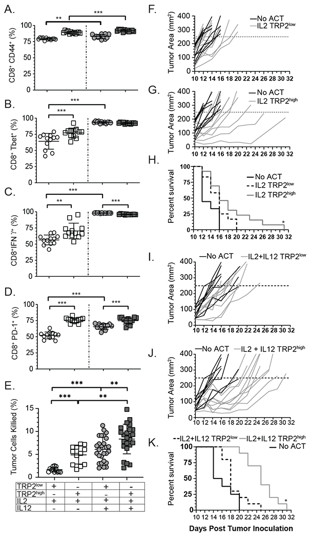
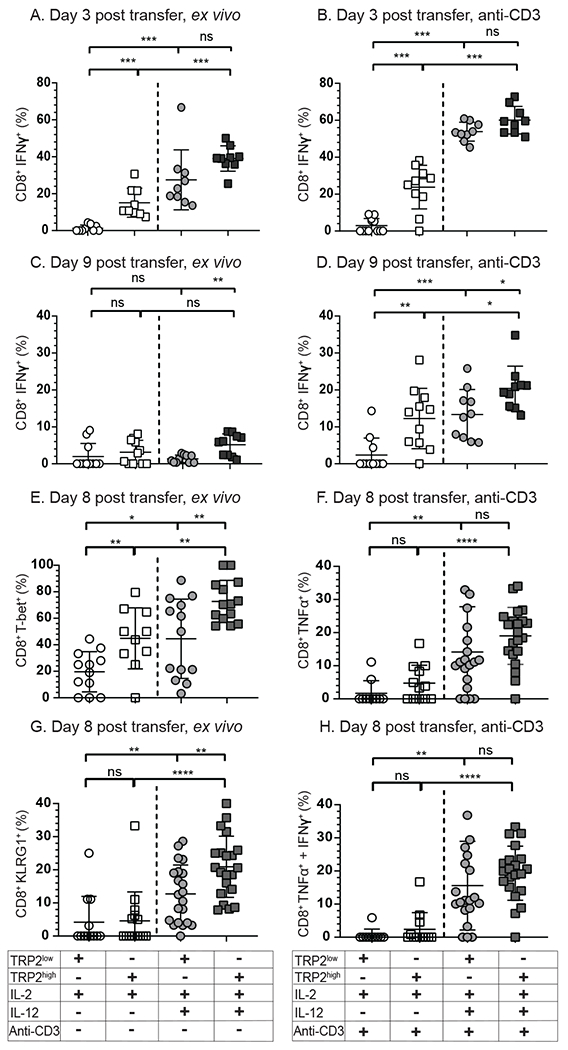
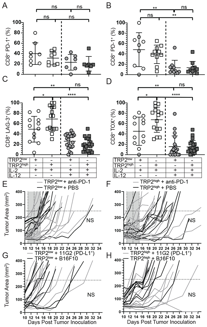
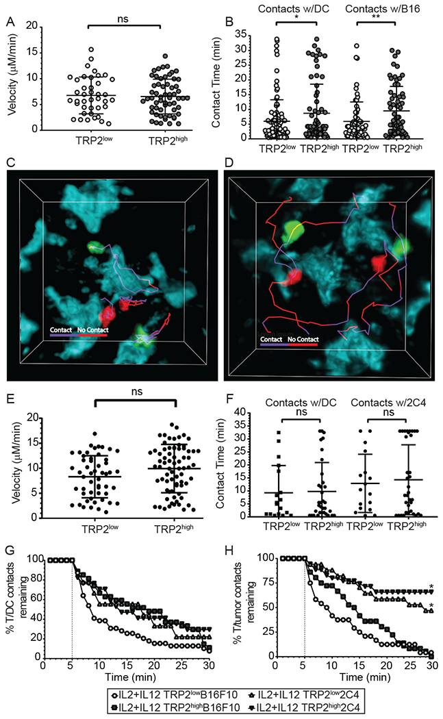
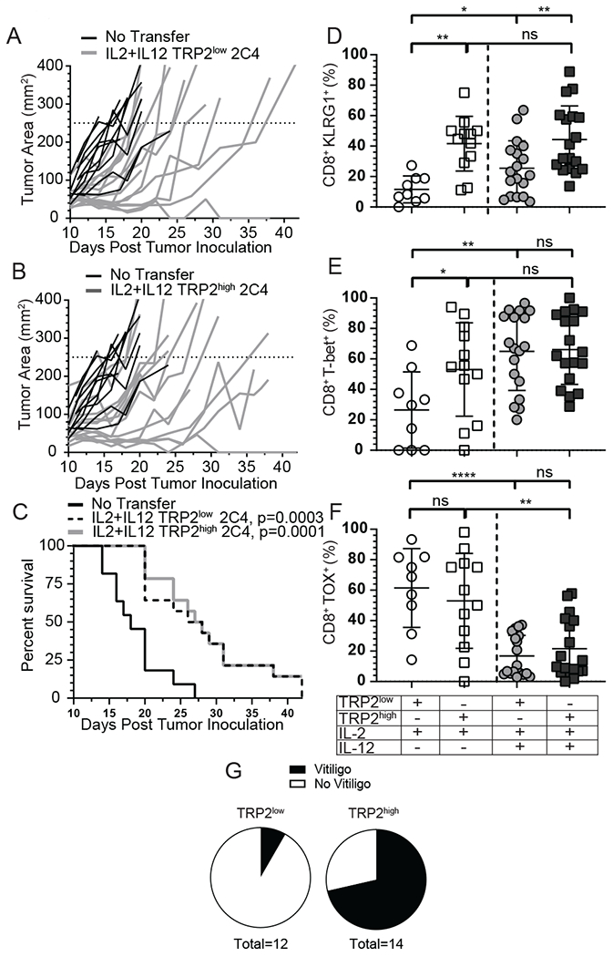
Optimal ex vivo expansion protocols of tumor-specific T cells followed by adoptive cell therapy must yield T cells able to home to tumors and effectively kill them. Our previous study demonstrated ex vivo activation in the presence of IL-12-induced optimal CD8+ T cell expansion and melanoma regression; however, adverse side effects, including autoimmunity, can occur. This may be due to transfer of high-avidity self-specific T cells. In this study, we compared mouse low- and high-avidity T cells targeting the tumor Ag tyrosinase-related protein 2 (TRP2). Not surprisingly, high-avidity T cells provide superior tumor control, yet low-avidity T cells can promote tumor regression. The addition of IL-12 during in vitro expansion boosts low-avidity T cell responsiveness, tumor regression, and prevents T cell exhaustion. In this study, we demonstrate that IL-12-primed T cells are resistant to PD-1/PD-L1-mediated suppression and retain effector function. Importantly, IL-12 preconditioning prevented exhaustion as LAG-3, PD-1, and TOX were decreased while simultaneously increasing KLRG1. Using intravital imaging, we also determined that high-avidity T cells have sustained contacts with intratumoral dendritic cells and tumor targets compared with low-avidity T cells. However, with Ag overexpression, this defect is overcome, and low-avidity T cells control tumor growth. Taken together, these data illustrate that low-avidity T cells can be therapeutically beneficial if cocultured with IL-12 cytokine during in vitro expansion and highly effective in vivo if Ag is not limiting. Clinically, low-avidity T cells provide a safer alternative to high-avidity, TCR-engineered T cells, as IL-12-primed, low-avidity T cells cause less autoimmune vitiligo.


Figures

Figure 1:
Figure 1:. IL-12-primed CD8+ T-cells have superior effector function and tumor killing capacity compared to IL-2-primed cells.
Naïve CD8+ T-cells were enriched from spleen and lymph nodes from TRP2low or TRP2high mice and cultured on plates coated with 50 μg/mL anti-CD3 and 0.8 μg/mL recombinant B7-1 and 5 IU/mL recombinant human IL-2 and/or 10 ng/mL IL-12 for three days. Cells were analyzed for (A) CD44, (B) T-bet, (C) IFNγ or (D) PD-1 by flow cytometry. (A-D) n=12 for each condition from one representative experiment. Similar results were observed in three independent experiments (E) Activated T-cells were co-cultured for 12 hours with B16F10 tumor target cells at a ratio of 5:1 (Effectors:Targets) to determine in vitro killing capacity. B16F10 target cells were analyzed for anti-active caspase-3. Compiled data from 6 independent experiments. (F) B16F10 tumor bearing mice were treated with 5x10⁶ IL-2 activated TRP2low (G) TRP2high cells , or (I) TRP2low , (J) TRP2high activated in the presence of IL-2 plus IL-12. Mice were followed for tumor growth and survival (H and K). Individual mice are shown in panels F, G, I, J, with the combined survival shown in panels H and K. Data are from two independent experiments. ns = not statistically different, **=p<0.001, ***=p<0.0001.
Figure 2:
Figure 2:. IL-12 priming prevents exhaustion in the tumor environment.
(A-D) IFNγ production by TRP2 reactive T-cells on (A,B) day 3 or (C, D) day 9 post transfer into tumor bearing mice. TRP2highUBQ-GFP T-cells and TRP2lowACT-tdTomato T-cells were isolated from the tumor environment and analyzed directly ex vivo for (A, B) IFNγ or (E) T-bet or (G) KLRG-1 or after 4 hour stimulation with plate bound anti-CD3 for (B, D) IFNγ, (F) TNFα, or dual expression of (H) TNFα and IFNγ. Culture conditions and subsequent ex vivo stimulation are shown in the table below the plots. Compiled data from three experimental replicates, n= 3-6 per condition. Ns = not statistically different, **=p<0.001, ***=p<0.0001.three isolated FACS experiments.
Figure 3:
Figure 3:. IL-12 co-culture prevents T-cell exhaustion which is not enhanced by PD-1 blockade.
(A-B) PD-1 expression on TRP2 Low and High T-cells isolated from the tumor from three independent experiments on either D2-3 (A) or D9 (B). (C) LAG-3 and (D) TOX expression in TRP2low and TRP2high T-cells isolated from the tumor on D9 post adoptive transfer. (E-F) Mice were treated with 1x10⁶ IL-2 + IL-12 primed TRP2low or TRP2high T-cells and then given 200μg of anti-PD1 or PBS control on D1, 3 and 5 post transfer. Mice were followed for tumor growth. (G-H) Mice were inoculated with B16F10 or PD-L1 overexpression (11G2) tumor cells and adoptively transferred with 1x10⁶ IL-2 + IL-12 primed T-cells and followed for tumor growth over time. Data are from two independent replicates. ns = not statistically different. **=p<0.001, ***=p<0.0001.
Figure 4:
Figure 4:. Antigen overexpression restores the ability of IL-12 primed low avidity T-cells to make sustained contacts with tumor targets.
(A-H) CD11c-YFP mice received B16F10-TFP or 2C4 tumors. Ten days after inoculation, mice received 5-10x106 IL-2 plus IL-12 TRP2low and TRP2high T-cells. 72 hours after adoptive T-cell transfer, mice were euthanized and tumors were removed and analyzed by two-photon microscopy. (A, B) T-cells were identified with Brainbow tdTomato (TRP2low) or UBC-GFP (TRP2high) expression and analyzed using Imaris software for average velocity and contact time in B16F10 tumors. (C, D) Representative images showing TRP2low and TRP2high T-cell contacts with tumor cell surfaces using tracks. Purple tracks represent sustained contact with the T-cell and tumor cell, while red tracks represent no-contact between T-cells and tumor targets at the timepoint on the track. Two photon microscopy was performed on ex vivo 2C4 tumors and (E) velocity and (F) contact time with dendritic cells or tumor targets. (G-H) Contact decay was analyzed between TRP2low or TRP2high and (E) YFP+ DCs (F) or TFP+ tumor targets. Contacts lasting less than five minutes were excluded from analysis. Data are from two independent replicates. ns = not statistically different, **=p<0.001, ***=p<0.0001.
Figure 5:
Figure 5:. Antigen overexpression ameliorates effector function differences of IL-12 primed low or high avidity T-cells.
(A-C) A murine TRP2-TFP tumor clone that expressed stable and high levels of TRP2-TFP (2C4) was inoculated into recipient mice and 1x10⁶ IL-2 + IL-12 primed TRP2low or TRP2high T-cells or PBS control (no T cell transfer) were adoptively transferred on D10 post tumor inoculation. Mice were followed for (A-B) tumor growth and (C) survival. Data from three independent experiments. (D-F) 2C4 tumors and spleen were harvested on D8 post adoptive T-cell transfer and lymphocytes were analyzed for (D) KLRG-1 and intracellular expression of (E) T-bet and (F) TOX by flow cytometery. Data are from three independent replicates. ns = not statistically different, **=p<0.001, ***=p<0.0001. (G) OT-1xUBC-GFP or WT B6 mice were given 5x10⁶ IL-2 plus IL-12 in vitro primed TRP2highGFP or TRP2lowtdtomato T-cells mice were followed for vitiligo induction. Incidence is shown 3 months after adoptive transfer. Data from two independent experiments pooled incidence.

References

    1. Wei SC, Duffy CR, and Allison JP, Fundamental Mechanisms of Immune Checkpoint Blockade Therapy. Cancer Discovery, 2018. 8(9): p. 1069–1086. - PubMed
    1. Topalian SL, Hodi FS, Brahmer JR, Gettinger SN, Smith DC, McDermott DF, Powderly JD, Sosman JA, Atkins MB, Leming PD, Spigel DR, Antonia SJ, Drilon A, Wolchok JD, Carvajal RD, McHenry MB, Hosein F, Harbison CT, Grosso JF, and Sznol M, Five-Year Survival and Correlates Among Patients With Advanced Melanoma, Renal Cell Carcinoma, or Non-Small Cell Lung Cancer Treated With Nivolumab. JAMA Oncol, 2019. - PMC - PubMed
    1. Tumeh PC, Harview CL, Yearley JH, Shintaku IP, Taylor EJ, Robert L, Chmielowski B, Spasic M, Henry G, Ciobanu V, West AN, Carmona M, Kivork C, Seja E, Cherry G, Gutierrez AJ, Grogan TR, Mateus C, Tomasic G, Glaspy JA, Emerson RO, Robins H, Pierce RH, Elashoff DA, Robert C, and Ribas A, PD-1 blockade induces responses by inhibiting adaptive immune resistance. Nature, 2014. 515(7528): p. 568–571. - PMC - PubMed
    1. Pauken KE, Sammons MA, Odorizzi PM, Manne S, Godec J, Khan O, Drake AM, Chen Z, Sen DR, Kurachi M, Barnitz RA, Bartman C, Bengsch B, Huang AC, Schenkel JM, Vahedi G, Haining WN, Berger SL, and Wherry EJ, Epigenetic stability of exhausted T cells limits durability of reinvigoration by PD-1 blockade. Science, 2016. 354(6316): p. 1160–1165. - PMC - PubMed
    1. Naidoo J, Page DB, Li BT, Connell LC, Schindler K, Lacouture ME, Postow MA, and Wolchok JD, Toxicities of the anti-PD-1 and anti-PD-L1 immune checkpoint antibodies. Ann Oncol, 2015. 26(12): p. 2375–2391. - PMC - PubMed

Publication types

MeSH terms