. 2020 Oct;27(10):934-941.
doi: 10.1038/s41594-020-0478-5. Epub 2020 Jul 31.

A thermostable, closed SARS-CoV-2 spike protein trimer

Collaborators, Affiliations

A thermostable, closed SARS-CoV-2 spike protein trimer

Xiaoli Xiong et al. Nat Struct Mol Biol. 2020 Oct.

Abstract

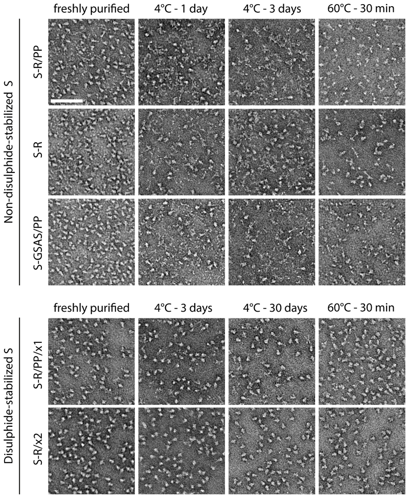
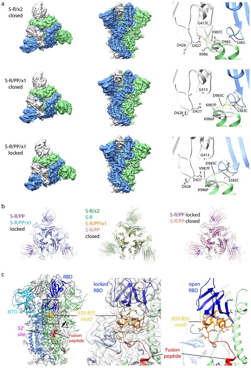
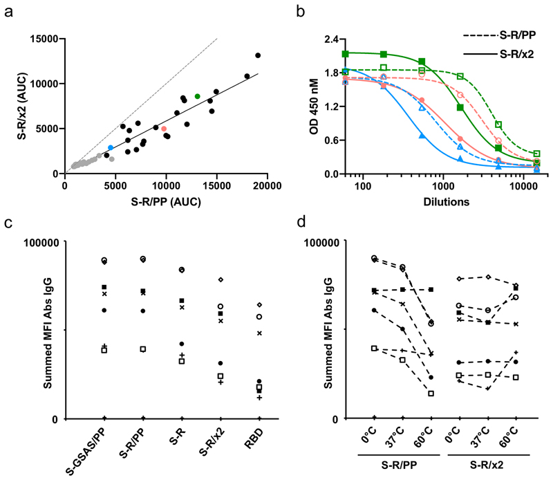
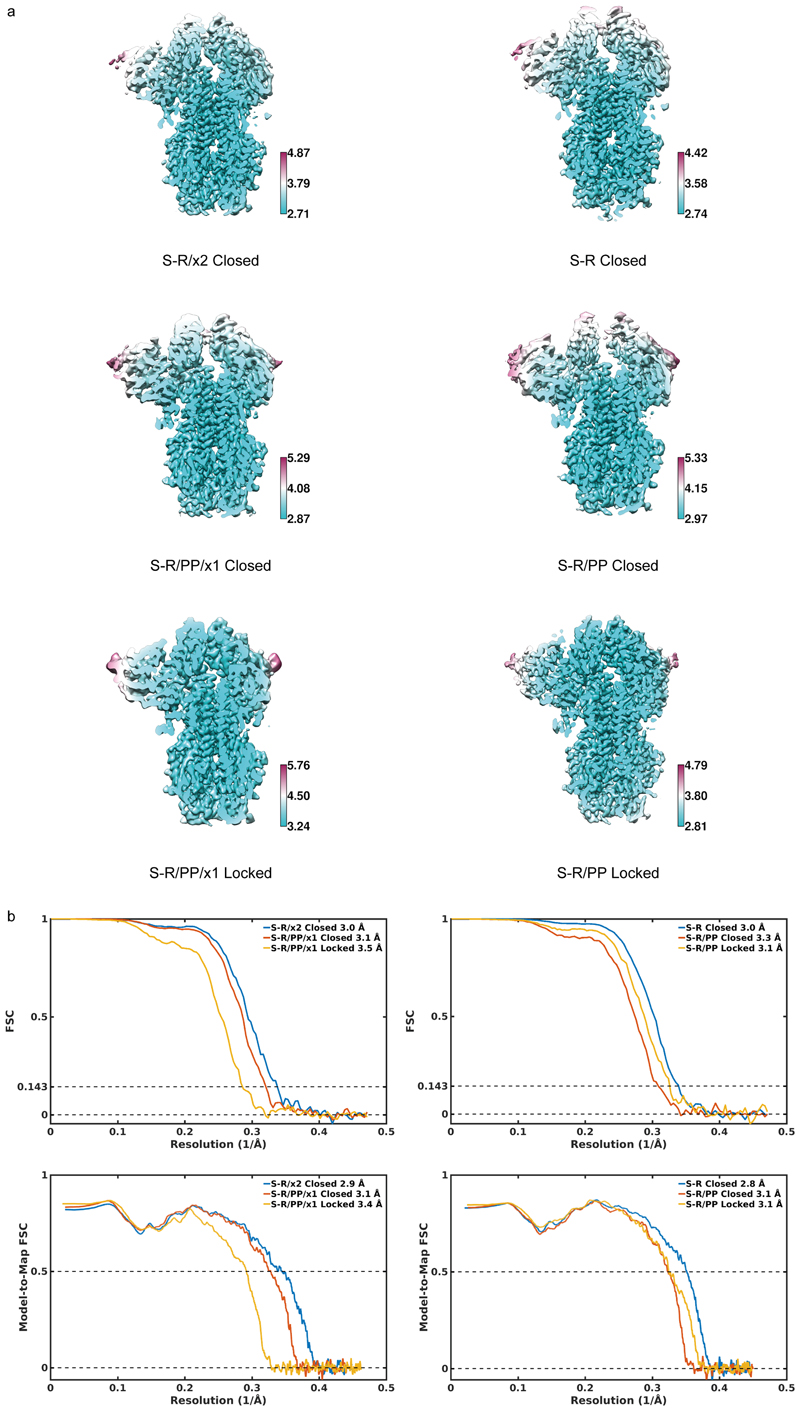
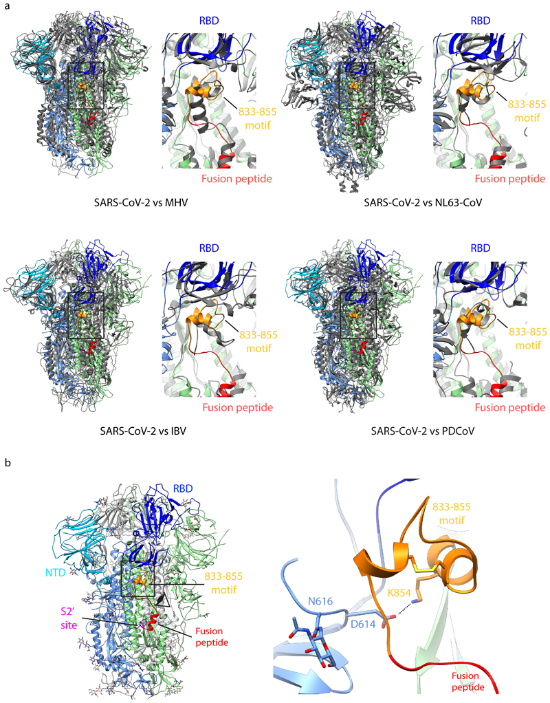
The spike (S) protein of SARS-CoV-2 mediates receptor binding and cell entry and is the dominant target of the immune system. It exhibits substantial conformational flexibility. It transitions from closed to open conformations to expose its receptor-binding site and, subsequently, from prefusion to postfusion conformations to mediate fusion of viral and cellular membranes. S-protein derivatives are components of vaccine candidates and diagnostic assays, as well as tools for research into the biology and immunology of SARS-CoV-2. Here we have designed mutations in S that allow the production of thermostable, disulfide-bonded S-protein trimers that are trapped in the closed, prefusion state. Structures of the disulfide-stabilized and non-disulfide-stabilized proteins reveal distinct closed and locked conformations of the S trimer. We demonstrate that the designed, thermostable, closed S trimer can be used in serological assays. This protein has potential applications as a reagent for serology, virology and as an immunogen.


Conflict of interest statement

Competing interests: The authors have no competing interests.

Figures

Figure 1
Figure 1. Design and expression of S protein constructs.
a) A schematic of the structure of trimeric S in its closed, prefusion form (PDB 6VXX) illustrating the location of the RBD and the NTD. Box indicates region shown in the right hand panel which illustrates the positions of disulphide bonds x1 and x2. Note that in PDB 6VXX and other published SARS-CoV-2 S protein structures, residues K986 and V987 have been mutated to proline. b) Western blots using antibodies against the C-terminal 6xHis tagged S-protein constructs in small-scale expression supernatants under non-reducing and reducing conditions. S-GSAS/PP/x2 and S-GSAS/x1 showed very low expression levels and were not considered further. c) Comparison of non-reducing and reducing Coomassie stained gels from medium-scale purifications of S-protein constructs. S-GSAS/PP/x1 and S-R/PP/x1 show a mixture of trimers and lower order oligomers under non-reducing conditions, whereas S-GSAS/x2 and S-R/x2 show trimers under these conditions.
Figure 2
Figure 2. Oligomeric state and thermal stability of S protein trimers assessed by negative stain EM.
Freshly purified proteins were imaged by negative stain EM (left column). Non-disulphide-stabilized S proteins show predominantly trimers with some smaller material, while S-R/x2 shows almost exclusively trimers. Upon prolonged incubation at 4°C, or upon heat treatment, non-disulphide-stabilized S proteins disintegrate, while disulphide-stabilized S proteins remain trimeric. Scale bar = 100 nm.
Figure 3
Figure 3. Characterisation of S-protein trimers by cryo-EM.
a) Cryo-EM densities for S-R/x2 in the closed conformation, and for S-R/PP/x1 in both closed and locked conformations. Boxes mark regions shown in right hand panels which illustrate resolved density for the inserted disulphide bonds at the expected positions. b) Comparison of the RBD and NTD conformations in S trimers of locked and closed conformations as viewed from the top of the trimer at the three-fold axis after alignment based on the central S2 helices. Between the locked and closed conformations the S1 domains show a twist motion (right panel and compare left and middle panel). In the locked conformation, the RBD moves closer to the 3-fold axis. The position of the NTD is variable. c) Folding of 833-855 into a helix-turn-helix motif locks the RBD in a locked conformation. Left panel, locations of RBD, 833-855 motif (orange), fusion peptide and S2' site are coloured and indicated. Middle panel, detailed density for the 833-855 motif is viewed from near the fusion peptide towards the RBD (indicated by the eye icon in the left panel). The density shows well resolved density that was disordered in previous SARS-CoV-1 and SARS-CoV-2 S structures. Right panel, the position of the RBD in the open conformation is aligned onto the locked monomer. In the open conformation the RBD would clash into the folded 833-855 motif (orange).
Figure 4
Figure 4. Behaviour of disulphide-stabilizedproteins in diagnostic assays.
a-b) Comparison of immune responses to open and closed conformations of the SARS-CoV-2 spike protein. Serum samples from PCR positive patients were screened for reactivity against S-R/PP and any samples scored as positive (n=48) were subsequently selected for end-point titrations against both for S-R/PP and S-R /x2. a). The area under the curve (AUC) was calculated for each seropositive sample using end point titrations. Paired AUC values for reactivity against S-R/PP and S-R /x2 were plotted for each positive sample. Pearson’s correlation analysis was calculated as r=0.9599 (95% CI 0.929 – 0.977) R2 of 0.921. The dashed line represents the theoretical line of equality, whereas the solid line represents the best fit using linear regression. Where possible, samples were used for subsequent EC50 determination (Extended Data Fig.4). Data points in grey are those where an EC50 for both S-R/PP and S-R /x2 could not be determined as one or both did not reach saturation. Coloured data points highlight three representative samples shown in panel b. b) End point titration curves against S-R/PP and S-R /x2. In each case the dashed line represents reactivity against the open conformation S-R/PP and the solid line represent the reactivity against the closed conformation S-R /x2. c-d) Summed mean fluorescence intensity (MFI) in a multiplexed flow cytometry (Luminex) assay to measure binding of IgG in sera to S protein variants and RBD. Sera from seven individuals seropositive for SARS-CoV-2 are shown, as well as two negative control sera (points along the x-axis). In d), S proteins were incubated at the indicated temperatures prior to use in the assay.
None
None
None
None

References

    1. Zhou P, et al. A pneumonia outbreak associated with a new coronavirus of probable bat origin. Nature. 2020;579:270–273. - PMC - PubMed
    1. Zhu N, et al. A Novel Coronavirus from Patients with Pneumonia in China, 2019. N Engl J Med. 2020;382:727–733. - PMC - PubMed
    1. Gorbalenya AE, et al. The species and its viruses – a statement of the Coronavirus Study Group. Biorxiv. 2020 doi: 10.1101/2020.02.07.937862. - DOI
    1. Li F. Structure, Function, and Evolution of Coronavirus Spike Proteins. Annu Rev Virol. 2016;3:237–261. - PMC - PubMed
    1. Graham BS, Gilman MSA, McLellan JS. Structure-Based Vaccine Antigen Design. Annu Rev Med. 2019;70:91–104. - PMC - PubMed

Publication types

MeSH terms