

Minimally invasive breast cancer excision using the breast lesion excision system under ultrasound guidance
- PMID: 32737712
- PMCID: PMC7568696
- DOI: 10.1007/s10549-020-05814-z
Minimally invasive breast cancer excision using the breast lesion excision system under ultrasound guidance
Abstract
Purpose: To assess the feasibility of completely excising small breast cancers using the automated, image-guided, single-pass radiofrequency-based breast lesion excision system (BLES) under ultrasound (US) guidance.
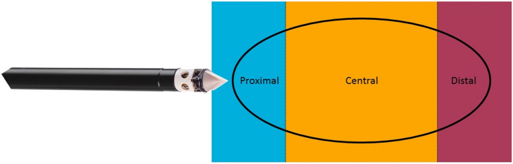
Methods: From February 2018 to July 2019, 22 patients diagnosed with invasive carcinomas ≤ 15 mm at US and mammography were enrolled in this prospective, multi-center, ethics board-approved study. Patients underwent breast MRI to verify lesion size. BLES-based excision and surgery were performed during the same procedure. Histopathology findings from the BLES procedure and surgery were compared, and total excision findings were assessed.
Results: Of the 22 patients, ten were excluded due to the lesion being > 15 mm and/or being multifocal at MRI, and one due to scheduling issues. The remaining 11 patients underwent BLES excision. Mean diameter of excised lesions at MRI was 11.8 mm (range 8.0-13.9 mm). BLES revealed ten (90.9%) invasive carcinomas of no special type, and one (9.1%) invasive lobular carcinoma. Histopathological results were identical for the needle biopsy, BLES, and surgical specimens for all lesions. None of the BLES excisions were adequate. Margins were usually compromised on both sides of the specimen, indicating that the excised volume was too small. Margin assessment was good for all BLES specimens. One technical complication occurred (retrieval of an empty BLES basket, specimen retrieved during subsequent surgery).
Conclusions: BLES allows accurate diagnosis of small invasive breast carcinomas. However, BLES cannot be considered as a therapeutic device for small invasive breast carcinomas due to not achieving adequate excision.
Keywords: Biopsy; Breast; Breast cancer; Minimally invasive; Vacuum.
Conflict of interest statement
The authors of this manuscript declare relationships with the following companies, whose products or services may be related to the subject matter of the article. In an associated clinical study, Medtronic the producer of the BLES needles has provided a research grant and non-financial support (BLES device and needles). W.B.G. Sanderink, L.J.A. Strobbe, P. Bult, M.S. Schlooz-Vries, S. Lardenoije, D. Venderink, and W. Vreuls declare no conflicts of interest. I. Sechopoulos has received research grants and research support from Siemens Healthineers, Canon Medical Systems and is scientific advisor of Fischer Medical. N. Karssemeijer is shareholder of Matakina Technology Limited Consultant, QView Medical, ScreenPoint Medical BV and is director of ScreenPoint Medical. R.M. Mann has received research grants and research support from Siemens Healthineers, Bayer Medical, Seno Medical, Elswood, Identification Solutions, Micrima and is Scientific advisor of Screenpoint Medical, Transonic Imaging.
Figures
References
-
- Verbeek ALM, Broeders MJM, Otto SJ, Fracheboud J, Otten JDMH, Holland R, Heeten GJAD, Koning HJD. Effecten van het bevolkingsonderzoek naar borstkanker. Ned Tijdschr Geneeskd. 2013;157:A5218. - PubMed
-
- Kaviani A, Sodagari N, Sheikhbahaei S, Eslami V, Hafezi-Nejad N, Safavi A, Noparast M, Fitoussi A. From radical mastectomy to breast-conserving therapy and oncoplastic breast surgery: a narrative review comparing oncological result, cosmetic outcome, quality of life, and health economy. ISRN Oncol. 2013;2013:742462. doi: 10.1155/2013/742462. - DOI - PMC - PubMed
MeSH terms
Grants and funding
LinkOut - more resources
Full Text Sources
Medical
