. 2020 Sep 9;28(3):486-496.e6.
doi: 10.1016/j.chom.2020.06.020. Epub 2020 Jul 3.

A Replication-Competent Vesicular Stomatitis Virus for Studies of SARS-CoV-2 Spike-Mediated Cell Entry and Its Inhibition

Affiliations

A Replication-Competent Vesicular Stomatitis Virus for Studies of SARS-CoV-2 Spike-Mediated Cell Entry and Its Inhibition

M Eugenia Dieterle et al. Cell Host Microbe. .

Abstract

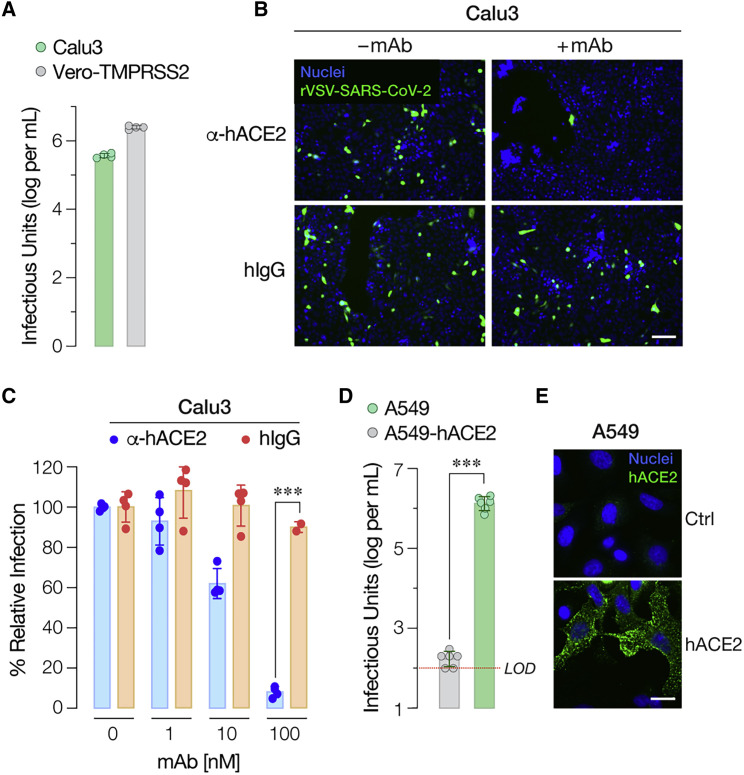
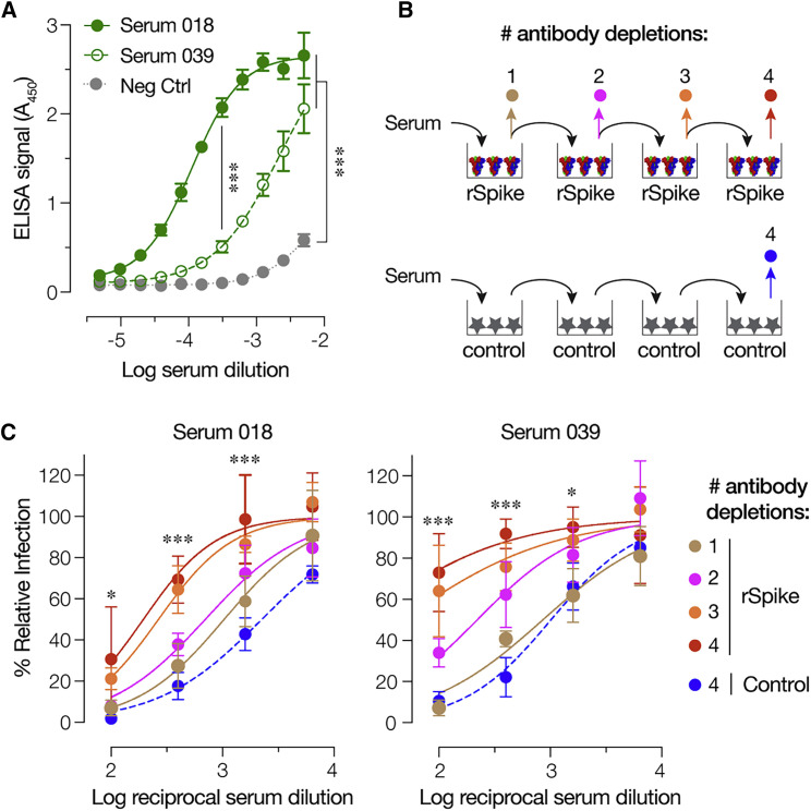
There is an urgent need for vaccines and therapeutics to prevent and treat COVID-19. Rapid SARS-CoV-2 countermeasure development is contingent on the availability of robust, scalable, and readily deployable surrogate viral assays to screen antiviral humoral responses, define correlates of immune protection, and down-select candidate antivirals. Here, we generate a highly infectious recombinant vesicular stomatitis virus (VSV) bearing the SARS-CoV-2 spike glycoprotein S as its sole entry glycoprotein and show that this recombinant virus, rVSV-SARS-CoV-2 S, closely resembles SARS-CoV-2 in its entry-related properties. The neutralizing activities of a large panel of COVID-19 convalescent sera can be assessed in a high-throughput fluorescent reporter assay with rVSV-SARS-CoV-2 S, and neutralization of rVSV-SARS-CoV-2 S and authentic SARS-CoV-2 by spike-specific antibodies in these antisera is highly correlated. Our findings underscore the utility of rVSV-SARS-CoV-2 S for the development of spike-specific therapeutics and for mechanistic studies of viral entry and its inhibition.

Keywords: ACE2; COVID-19; SARS-CoV-2; VSV; antiviral drugs; convalescent plasma; neutralization assay; neutralizing antibody; serology; surrogate.


Conflict of interest statement

Declaration of Interests K.C. is a member of the scientific advisory board of Integrum Scientific, LLC.

Figures

None
Graphical abstract
Figure 1
Figure 1
Generation of a Recombinant Vesicular Stomatitis Virus (rVSV) Bearing the SARS-CoV-2 Spike (S) Glycoprotein (A) Schematic representation of the VSV genome in which its native glycoprotein gene has been replaced by that encoding the SARS-CoV-2 S protein. The VSV genome has been further modified to encode an enhanced green fluorescent protein (eGFP) reporter to easily score for infection. (B) Infectious center formation assay on Vero cells at 24 h post-infection showing growth of the rVSV-SARS-CoV-2 S after the indicated number of rounds of serial passage of the passage #1 virus (carrying wild-type [WT] S sequences) on Huh7.5.1 cell line (scale bar, 100 μm). Two representative images for each virus passage, showing infected cells pseudo-colored in green, from one of the two independent experiments are shown here. (C) Incorporation of SARS-CoV-2 S into rVSV particles captured on an ELISA plate was detected using antiserum from a COVID-19 convalescent donor (average ± SD, n = 12 from 3–4 independent experiments). Serum from a COVID-19-negative donor and rVSVs bearing Ebola virus glycoprotein (EBOV GP) were used as negative controls (average ± SD, n = 6 from 2 independent experiments). (D) Representative images showing Vero cells infected with plaque #2, #3, and #6 viruses at 16 h post-infection (scale bar, 100 μm). (E) Production of infectious virions at 48 h post-infection from Vero cells infected with the indicated plaque-purified viruses. Titers were measured on Vero cells overexpressing TMPRSS2 (n = 4, from two independent titrations).
Figure 2
Figure 2
rVSV-SARS-CoV-2 S Infection Requires the Activity of Cysteine Cathepsin Proteases (A) Huh7.5.1 cells pre-treated for 1 h at 37°C with the indicated concentrations of NH4Cl were infected with pre-titrated amounts of rVSVs bearing SARS-CoV-2 S or EBOV GP. Infection was scored by eGFP expression at 16–18 h post-infection (average ± SD, n = 8 from 2 independent experiments). (B) Vero cells pre-treated for 90 min at 37°C with the indicated concentrations of pan-cysteine cathepsin inhibitor E-64 were infected with pre-titrated amounts of rVSVs bearing SARS-CoV-2 S, EBOV GP, or VSV G and scored for infection as above (average ± SD, n = 6 from 3 independent experiments, except n = 4 from 2 independent experiments for EBOV GP). (C) Vero cells pre-treated for 90 min at 37°C with the indicated concentrations of cathepsin L/B inhibitor FYdmk were infected with pre-titrated amounts of rVSVs bearing SARS-CoV-2 S, EBOV GP, or VSV G. Infection was scored as above (average ± SD, n = 6 from 3 independent experiments). (D) Vero cells and Vero cells overexpressing TMPRSS2 pre-treated for 120min at 37C with the indicated concentrations of camostat ]were infected with pre-titrated amounts of rVSVs bearing SARS-CoV-2 S and subsequently scored for infection. In panels (B)–(D), all comparisons were made between vehicle- and inhibitor-treated samples. ns,not statistically significant. p < 0.033, ∗∗∗p < 0.001.
Figure 3
Figure 3
rVSV-SARS-CoV-2 S Infection Requires Human ACE2 (A) Naive (None) baby hamster kidney (BHK21) cells or cells transduced with a retrovirus carrying human ACE2 cDNA (+ hACE2 cDNA) were immunostained for hACE2 expression using an anti-ACE2 antibody. Cells were imaged by fluorescence microscopy. The hACE2 signal is pseudo-colored green (top panel, scale bar = 20 μm). These cells were also exposed to serial 5-fold dilutions of rVSV-SARS-CoV-2 S and infection was scored by eGFP expression (bottom panel, scale bar = 50 μm). Representative images from one of 3 independent experiments are shown. (B) Enumeration of eGFP-positive green cells (Average ± SD, n = 8 from 3 independent experiments). Red dotted line indicates the assay limit of detection (LOD). (C) Recombinant, Ni-NTA–affinity purified S1-S2 ectodomain (Spike) or the receptor binding domain (RBD) of the SARS-CoV-2 S protein were subjected to SDS-PAGE and Coomassie staining. A representative image from one of two independent purification trials is shown here. (D) Monolayers of Huh7.5.1 cells were pre-incubated with serial 3-fold dilutions of the purified RBD for 1 h at 37°C and then infected with pre-titrated amounts of rVSVs bearing SARS-CoV-2 S or EBOV GP. At 16–18 h post-infection, cells were fixed, nuclei counter-stained with Hoechst-33342, and infection (eGFP expression) was scored by fluorescence microscopy. It is represented as % relative infection (no RBD = 100%, Average ± SEM, n = 8 from 3–4 [rVSV-SARS-CoV-2 S] or n = 4 from 2 [rVSV-EBOV GP] independent experiments). (E) Monolayers of Huh7.5.1 cells pre-incubated for 1 h at 37°C with 3-fold serial dilutions of anti-human ACE2 antibody or negative control (hIgG) were infected with pre-titrated amounts of rVSV-SARS-CoV-2 S. Infection was scored as above and is represented as % relative infection (no antibody = 100%, Average ± SD, n = 8 from 3–4 independent experiments). ∗∗∗p < 0.001.
Figure 4
Figure 4
rVSV-SARS-CoV-2 S Infection in Human Airway Epithelial Cells is ACE2-Dependent (A) Infectivity of rVSV-SARS-CoV-2 S was measured in human airway epithelial Calu3 cells and Vero-TMPRSS2 cells by applying serial dilutions of the virus. Infections were scored as described in Figure 3B (Average ± SD, n = 4 from two independent titrations). (B and C) Monolayers of Calu3 cells pre-incubated for 1 h at 37°C with indicated amounts of anti-human ACE2 antibody or negative control (hIgG) were infected with pre-titrated amounts of rVSV-SARS-CoV-2 S. (B) Representative images from one of the two independent experiments are shown (scale bar, 100 μm). (C) Infection was scored as above and is represented as % relative infection (no antibody = 100%, Average ± SD, n = 4 from two independent experiments, except for n = 2 for hIgG at 100 nM). (D) Infectivity of rVSV-SARS-CoV-2 S in human respiratory epithelial A549 cells transduced with a retrovirus carrying human ACE2 cDNA or empty vector was evaluated by exposing cells to serial dilutions of rVSV-SARS-CoV-2 S. Infections were scored as described in Figure 3B (Average ± SD, n = 6 from 3 independent titrations). Red dotted line indicates the assay limit of detection (LOD). Means were compared by unpaired t test. (E) A549 cells transduced as in panel 3D were immunostained for hACE2 expression as described in Figure 3A. using an anti-ACE2 antibody. Cells were imaged by fluorescence microscopy. The hACE2 signal is pseudo-colored green and representative images are shown (scale bar = 20 μm). ∗∗∗p < 0.001.
Figure 5
Figure 5
rVSV-SARS-CoV-2 S Neutralization Is Mediated by S-Glycoprotein-Targeting Antibodies in Human Antisera (A) ELISA plates coated with rVSV-SARS-CoV-2 S were incubated with serial 2-fold dilutions of serum 18, serum 39, or negative control serum. Bound S-specific antibodies were detected with an anti-human HRP-conjugated secondary antibody (average ± SD, n = 4 from 2 independent experiments). (B) Schematic of the antibody depletion study. (C) Pre-titrated amounts of serum 18 and serum 39 were sequentially incubated with SARS-CoV-2 S-coated high-binding plates to deplete S-specific antibodies. Capacity of the depleted sera (and control sera incubated with only the blocking agent) to neutralize rVSV-SARS-CoV-2 S was then estimated by incubating pre-titrated amounts of rVSV at the indicated dilutions of sera at 37°C for 1 h prior to infecting monolayers of Huh7.5.1 cells. Cells were scored for infection as above (average ± SD, n = 4 from 2 independent experiments). In panels (A), (C), and (D), p values for pairwise comparisons of the untreated sample and inhibitor-treated sample means are shown, unless otherwise indicated. In panels (C) and (D), the depletion #4 and depletion control were compared. p < 0.033,∗∗∗p < 0.001.
Figure 6
Figure 6
Correlation of Convalescent Serum-Mediated Neutralization of rVSV-SARS-CoV-2 S and Authentic SARS-CoV-2 (A) Pre-titrated amounts of SARS-CoV-2 were incubated with serial 3-fold dilutions of antisera from COVID-19 convalescent donors or negative control at 37°C for 1 h. Virus-serum mixtures were then applied to monolayers of Vero-E6 cells. At 24 h post-infection, cells were fixed, permeabilized and immunostained with a SARS-CoV nucleocapsid-specific antibody. Nuclei were counterstained, and infected cells were scored for the presence of nucleocapsid antigen. Representative images from one of the 2 independent experiments are shown (scale bar, 200 μm). (B) Pre-titrated amounts of rVSV-SARS-CoV-2 S were incubated with serial 3-fold dilutions of antisera from COVID-19 convalescent patients or negative control at 37°C for 1 h. Virus:serum mixtures were then applied to monolayers of Vero cells. At 7 h post-infection, cells were fixed, nuclei were counterstained, and infected cells were scored by GFP expression. Heatmaps showing % neutralization of authentic SARS-CoV-2 or rVSV-SARS-CoV-2 S by the panel of 40 antisera are shown (Averages of n = 4 from 2 independent experiments). (C and D) Comparison of the neutralizing activities of the antisera (log reciprocal IC50 values) against authentic SARS-CoV-2 and rVSV-SARS-CoV-2 S (“rVSV SARS2 S”). (D) Linear regression analyses of neutralization IC50 values from panel C.

Update of

Comment in

  • Snatching the Crown from SARS-CoV-2.
    Coughlan L. Coughlan L. Cell Host Microbe. 2020 Sep 9;28(3):360-363. doi: 10.1016/j.chom.2020.08.007. Cell Host Microbe. 2020. PMID: 32910919 Free PMC article.

References

    1. Amanat F., Stadlbauer D., Strohmeier S., Nguyen T.H.O., Chromikova V., McMahon M., Jiang K., Arunkumar G.A., Jurczyszak D., Polanco J. A serological assay to detect SARS-CoV-2 seroconversion in humans. Nat. Med. 2020 doi: 10.1038/s41591-020-0913-5. - DOI - PMC - PubMed
    1. Bestle D., Heindl M.R., Limburg H., Van Lam van T., Pilgram O., Moulton H., Stein D.A., Hardes K., Eickmann M., Dolnik O. TMPRSS2 and furin are both essential for proteolytic activation and spread of SARS-CoV-2 in human airway epithelial cells and provide promising drug targets. bioRxiv. 2020 doi: 10.1101/2020.04.15.042085. - DOI - PMC - PubMed
    1. Bosch B.J., van der Zee R., de Haan C.A.M., Rottier P.J.M. The coronavirus spike protein is a class I virus fusion protein: structural and functional characterization of the fusion core complex. J. Virol. 2003;77:8801–8811. - PMC - PubMed
    1. Böttcher-Friebertshäuser E., Freuer C., Sielaff F., Schmidt S., Eickmann M., Uhlendorff J., Steinmetzer T., Klenk H.-D., Garten W. Cleavage of influenza virus hemagglutinin by airway proteases TMPRSS2 and HAT differs in subcellular localization and susceptibility to protease inhibitors. J. Virol. 2010;84:5605–5614. - PMC - PubMed
    1. Böttcher-Friebertshäuser E., Garten W., Matrosovich M., Klenk H.D. The hemagglutinin: a determinant of pathogenicity. Curr. Top. Microbiol. Immunol. 2014;385:3–34. - PubMed

Publication types

MeSH terms