. 2020 Sep 3;79(5):768-781.e7.
doi: 10.1016/j.molcel.2020.07.009. Epub 2020 Jul 31.

Quality Control of ER Membrane Proteins by the RNF185/Membralin Ubiquitin Ligase Complex

Affiliations

Quality Control of ER Membrane Proteins by the RNF185/Membralin Ubiquitin Ligase Complex

Michael L van de Weijer et al. Mol Cell. .

Erratum in

Abstract

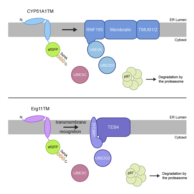
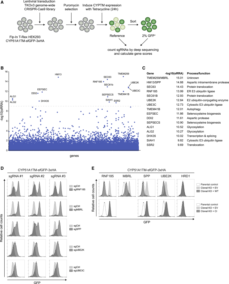
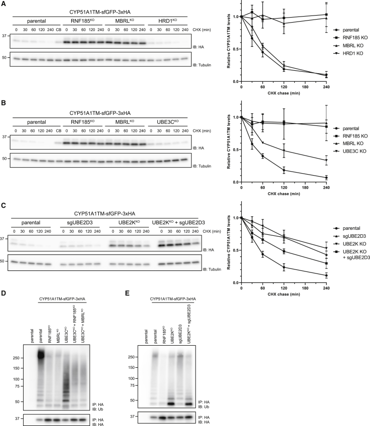
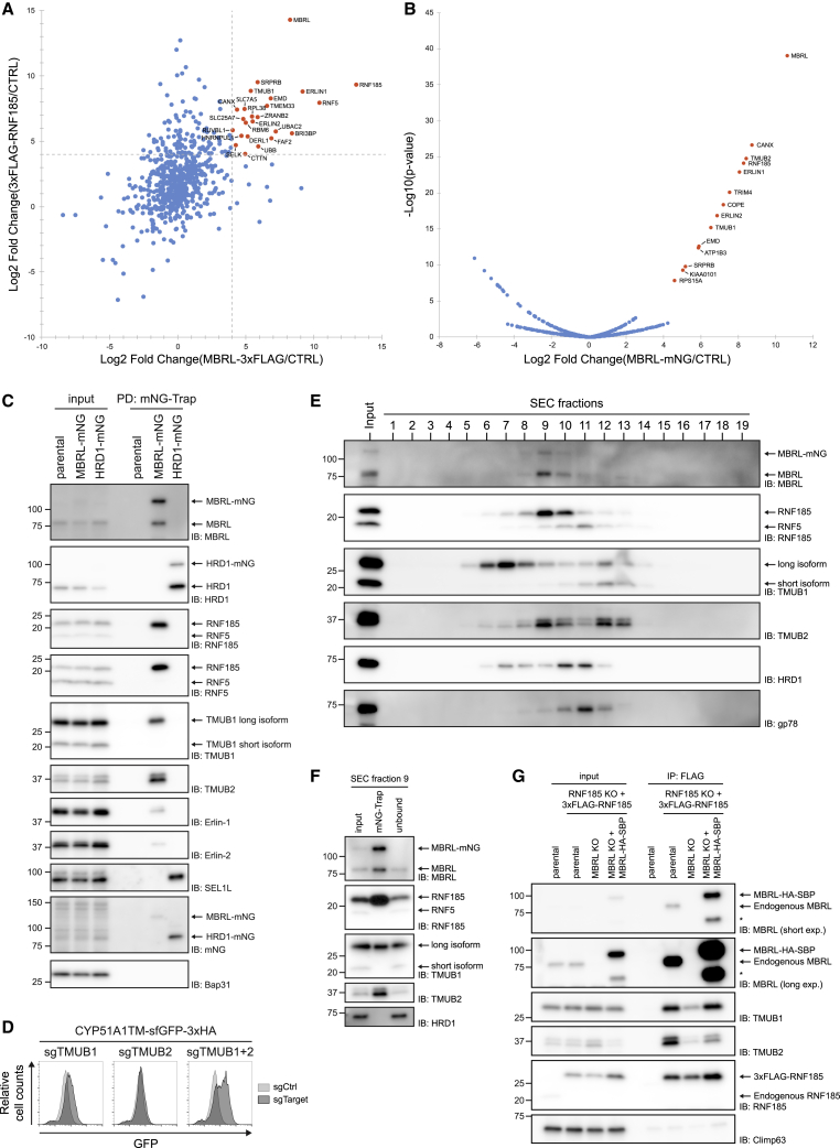
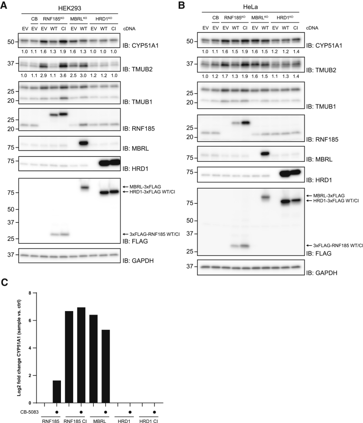
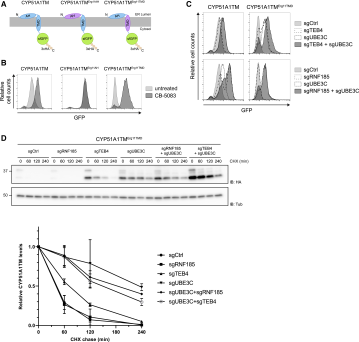
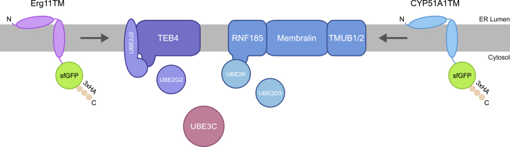
Misfolded proteins in the endoplasmic reticulum (ER) are degraded by ER-associated degradation (ERAD). Although ERAD components involved in degradation of luminal substrates are well characterized, much less is known about quality control of membrane proteins. Here, we analyzed the degradation pathways of two short-lived ER membrane model proteins in mammalian cells. Using a CRISPR-Cas9 genome-wide library screen, we identified an ERAD branch required for quality control of a subset of membrane proteins. Using biochemical and mass spectrometry approaches, we showed that this ERAD branch is defined by an ER membrane complex consisting of the ubiquitin ligase RNF185, the ubiquitin-like domain containing proteins TMUB1/2 and TMEM259/Membralin, a poorly characterized protein. This complex cooperates with cytosolic ubiquitin ligase UBE3C and p97 ATPase in degrading their membrane substrates. Our data reveal that ERAD branches have remarkable specificity for their membrane substrates, suggesting that multiple, perhaps combinatorial, determinants are involved in substrate selection.

Keywords: ER-associated degradation; ERAD; RNF185; TEB4/MARCH6; TMEM259; TMUB1/TMUB2; UBE3C; endoplasmic reticulum; membralin; protein quality control.


Conflict of interest statement

Declaration of Interests The authors declare no competing interests.

Figures

None
Graphical abstract
Figure 1
Figure 1
Erg11TM and CYP51A1TM Engage Distinct ERAD Branches (A) Schematic representation of the Erg11TM and CYP51A1TM constructs. Erg11TM and CYP51A1TM were C-terminally tagged with sfGFP and a 3xHA-tag. The Erg11TM and CYP51A1TM amphipathic helix (AH) and transmembrane (TMD) domain are aligned and highlighted in red. (B) Subcellular fractionation of Erg11TM- and CYP51A1TM-expressing cells. Postnuclear supernatants (W) were fractionated into a cytosolic soluble fraction (C) and a crude membrane pellet (P1). Membranes in P1 were salt washed and carbonate treated (P2) to remove all peripherally associated proteins. Fractions were subjected to SDS-PAGE and western blotting analysis. The partitioning of the HA-tagged substrates was compared to endogenous soluble (p97 and GAPDH) and integral membrane (BAP31) proteins. (C and D) Erg11TM and CYP51A1TM levels are stabilized upon inhibition of p97 and the proteasome but are unaffected by blocking protein delivery to lysosomes. Flow cytometry (C) and immunoblotting (D) analysis of tetracycline-induced expression of Erg11TM and CYP51A1TM in Flp-In T-REx 293 cells. Analysis was performed 24 h post-induction in cells left untreated, incubated 4 h with inhibitors to p97 (CB-5083; 2.5 μM [CB]) or to the proteasome (Bortezomib; 500 nM [Btz]), or incubated 24 h with bafilomycin A (250 nM [Baf]) that inhibits lysosomal delivery. Flp-In T-REx 293 cells without any GFP-tagged substrate were used as control (no substrate). In (D), Erg11TM and CYP51A1TM were detected with anti-HA antibodies. LC3 was analyzed to confirm effectiveness of bafilomycin A treatment and was detected with an anti-LC3 antibody. Tubulin was used as a loading control and was detected with an anti-tubulin antibody. (E) Degradation of Erg11TM and CYP51A1TM was analyzed after inhibition of protein synthesis by cycloheximide (CHX) in the absence or presence of the p97 inhibitor CB-5083 (4 h; 2.5 μM). Cell extracts were analyzed by SDS-PAGE and immunoblotting. Erg11TM and CYP51A1TM were detected with anti-HA antibodies. GAPDH was used as a loading control and detected with an anti-GAPDH antibody. (F) Erg11TM and CYP51A1TM follow distinct ERAD pathways. Cells expressing Erg11TM and CYP51A1TM as well as plasmids encoding sgRNAs targeting the indicated genes were analyzed by flow cytometry (based on GFP fluorescence). (G) Degradation of Erg11TM depends on TEB4, UBE2G2, and UBE2J2. Erg11TM levels were analyzed by flow cytometry (based on GFP fluorescence) in parental control cells and in clonal TEB4, UBE2G2, UBE2J2, and HRD1 KO cells expressing cDNAs encoding either the corresponding wild type (WT), a catalytically inactive (CI) mutant, or an empty vector (EV). See also Figure S1.
Figure 2
Figure 2
A Genome-wide CRISPR-Cas9 Screen Identifies Components Required for CYP51A1TM Degradation (A) Workflow of the CRISPR-Cas9 genome-wide screen. (B) Significance score of the genes analyzed in the screen calculated by the MAGeCK algorithm. The x axis represents the genes in alphabetical order. The y axis shows the −log(αRRA) significance value. The −log(αRRA) cutoff was arbitrarily set at 9 (dashed line). Significantly enriched genes are annotated. (C) Overview of the enriched genes identified and their proposed function. (D) Validation of several screen hits using independent sgRNAs. Levels of CYP51A1TM were analyzed by flow cytometry (based on GFP fluorescence). (E) Degradation of CYP51A1TM depends on RNF185, MBRL, SPP, and UBE2K. CYP51A1TM levels were assessed by flow cytometry (based on GFP fluorescence) in parental control cells and clonal RNF185, MBRL, SPP, UBE2K, and HRD1 KO cells expressing cDNAs encoding either WT, a CI mutant, or an EV. See also Figure S2.
Figure 3
Figure 3
CYP51A1TM Ubiquitination and Degradation Are Dependent on RNF185 and Membralin (A) RNF185 and Membralin are essential for CYP51A1TM degradation. Degradation of CYP51A1TM was analyzed upon inhibition of protein synthesis with cycloheximide (CHX) in parental, RNF185, MBRL, and HRD1 KO cells. Cell extracts were analyzed by SDS-PAGE and immunoblotting. The graph shows the average of three experiments; error bars represent the standard deviation. (B) UBE3C promotes efficient CYP51A1TM degradation. Degradation of CYP51A1TM in cells lacking the indicated genes was analyzed as in (A). (C) The E2s UBE2K and UBE2D3 are involved in CYP51A1TM degradation. Degradation of CYP51A1TM in cells depleted for the indicated genes was analyzed as in (A). (D) Ubiquitination of CYP51A1TM is dependent on RNF185 and MBRL. CYP51A1TM was immunoprecipitated from parental cells or cells lacking the indicated genes and analyzed by SDS-PAGE followed by immunoblotting with anti-HA and anti-ubiquitin antibodies. Parental cells lacking CYP51A1TM substrate were used as a negative control. (E) UBE2K facilitates polyubiquitination of CYP51A1TM. CYP51A1TM ubiquitination in cells depleted for the indicated E2 enzymes was analyzed as in (D). See also Figure S3.
Figure 4
Figure 4
RNF185 and Membralin Form a Novel ERAD Complex (A) Proteins co-precipitating with MBRL-3xFLAG (x axis) and 3xFLAG-RNF185 (y axis) as analyzed by mass spectrometry. The enrichment of proteins associated to FLAG-tagged baits over an untagged control was used to calculate the log2 fold changes. Proteins enriched above an arbitrary cutoff of 4 in both MBRL and RNF185 IPs are annotated (in orange). (B) Proteins co-precipitating with endogenous Membralin-mNG as detected by mass spectrometry. The x axis shows the log2 fold change of MBRL-mNG versus untagged control cell line; the y axis shows the −log10 p value estimated by the SignificanceB analysis (Cox and Mann, 2008). Cutoff was arbitrarily set to a log2 (fold change) of 4 with a −log10 (p) of 5. Significantly enriched proteins are annotated (in orange). (C) Western blotting validation of the interactions identified in (B) by mNG-Trap pull-down (PD) of endogenously tagged Membralin-mNG. Endogenously tagged HRD1-mNG was used as specificity control. (D) Analysis of CYP51A1TM levels in cells depleted for TMUB1, TMUB2, or both. CYP51A1TM was analyzed by flow cytometry (based on GFP fluorescence). (E) Size exclusion chromatography of detergent-solubilized membranes of HeLa cells containing endogenously tagged MBRL-mNG (MBRL-mNG). Crude membranes were solubilized in 1% DDM + 0.1% CHS, and solubilized material was applied to a Superose 6 10/300 GL column. Elution fractions were analyzed by SDS-PAGE and western blotting with the indicated antibodies. (F) Fraction 9 from (E) was subjected to immunoprecipitation using mNG-Trap beads. Eluted material was analyzed by western blotting for the proteins indicated. (G) Membralin mediates the interaction between RNF185 and TMUBs. Immunoprecipitation of 3xFLAG-RNF185 in cells with the indicated gene deletions is shown. Eluted proteins were analyzed by SDS-PAGE and immunoblotting for the proteins indicated. The asterisk () indicates a truncated MBRL product. See also Figure S4.
Figure 5
Figure 5
Levels of Endogenous CYP51A1 and TMUB2 Are Regulated by the RNF185-MBRL Complex (A and B) Protein extracts from RNF185, MBRL, and HRD1 KO (A) HEK293 and (B) HeLa cells expressing either an EV or cDNAs encoding the WT or CI version of the indicated protein were analyzed by SDS-PAGE and immunoblotting. Extracts from parental cells left untreated or treated with the p97 inhibitor CB-5083 (CB) (4 h; 2.5 μM) were also analyzed. Relative CYP51A1 and TMUB2 levels are displayed below the respective blot, normalized to the GAPDH loading control. (C) Endogenous CYP51A1 co-precipitates specifically with RNF185/MBRL complex. The indicated FLAG-tagged proteins (x axis) were precipitated from untreated or CB-5083-treated cells, and eluted proteins were analyzed by mass spectrometry. The y axis shows the log2 fold enrichment of endogenous CYP51A1 in the FLAG precipitates versus untagged control. See also Figure S5.
Figure 6
Figure 6
RNF185-MBRL and TEB4 Recognize Distinct Features on Their Membrane Substrates (A) Schematic overview of the CYP51A1TM chimeras. The AH and the TMD from the Erg11TM ERAD substrate are indicated in purple. (B) The levels of CYP51A1TM chimeras were analyzed by flow cytometry (based on GFP fluorescence) in the absence or presence of the p97 inhibitor CB-5083 (4 h; 2.5 μM). (C) The levels of CYP51A1TM and the chimera CYP51A1TMErg11TMD were analyzed by flow cytometry (based on GFP fluorescence) in cells depleted for the indicated genes. (D) Degradation of CYP51A1TMErg11TMD analyzed upon inhibition of protein synthesis with CHX in cells depleted for the indicated genes. Cell extracts were analyzed by SDS-PAGE and immunoblotting. The graph shows the average of three experiments; error bars represent the standard deviation. See also Figure S6.
Figure 7
Figure 7
Erg11TM and CYP51A1TM Degradation by the TEB4 and RNF185-MBRL Ubiquitin Ligase Complex Schematic overview of Erg11TM and CYP51A1TM degradation by the TEB4 and RNF185-MBRL ubiquitin ligase complex, respectively.

References

    1. Andersson O., von Euler G. Characterization and expression of the gene encoding membralin, an evolutionary conserved protein expressed in the central nervous system. Brain Res. Gene Expr. Patterns. 2002;1:205–212. - PubMed
    1. Bernasconi R., Galli C., Calanca V., Nakajima T., Molinari M. Stringent requirement for HRD1, SEL1L, and OS-9/XTP3-B for disposal of ERAD-LS substrates. J. Cell Biol. 2010;188:223–235. - PMC - PubMed
    1. Carvalho P., Goder V., Rapoport T.A. Distinct ubiquitin-ligase complexes define convergent pathways for the degradation of ER proteins. Cell. 2006;126:361–373. - PubMed
    1. Chen Z., Pickart C.M. A 25-kilodalton ubiquitin carrier protein (E2) catalyzes multi-ubiquitin chain synthesis via lysine 48 of ubiquitin. J. Biol. Chem. 1990;265:21835–21842. - PubMed
    1. Chen Y.-C., Davidson B., Cheng C.-C., Maitra A., Giuntoli R.L., 2nd, Hruban R.H., Wang T.-L., Shih IeM. Identification and characterization of membralin, a novel tumor-associated gene, in ovarian carcinoma. Biochim. Biophys. Acta. 2005;1730:96–102. - PubMed

Publication types