. 2020 Sep:181:104882.
doi: 10.1016/j.antiviral.2020.104882. Epub 2020 Jul 29.

An enzyme-based immunodetection assay to quantify SARS-CoV-2 infection

Affiliations

An enzyme-based immunodetection assay to quantify SARS-CoV-2 infection

Carina Conzelmann et al. Antiviral Res. 2020 Sep.

Abstract

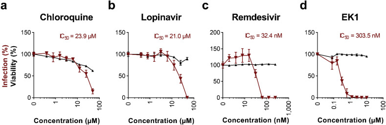
SARS-CoV-2 is a novel pandemic coronavirus that caused a global health and economic crisis. The development of efficient drugs and vaccines against COVID-19 requires detailed knowledge about SARS-CoV-2 biology. Several techniques to detect SARS-CoV-2 infection have been established, mainly based on counting infected cells by staining plaques or foci, or by quantifying the viral genome by PCR. These methods are laborious, time-consuming and expensive and therefore not suitable for a high sample throughput or rapid diagnostics. We here report a novel enzyme-based immunodetection assay that directly quantifies the amount of de novo synthesized viral spike protein within fixed and permeabilized cells. This in-cell ELISA enables a rapid and quantitative detection of SARS-CoV-2 infection in microtiter format, regardless of the virus isolate or target cell culture. It follows the established method of performing ELISA assays and does not require expensive instrumentation. Utilization of the in-cell ELISA allows to e.g. determine TCID50 of virus stocks, antiviral efficiencies (IC50 values) of drugs or neutralizing activity of sera. Thus, the in-cell spike ELISA represents a promising alternative to study SARS-CoV-2 infection and inhibition and may facilitate future research.

Trial registration: ClinicalTrials.gov NCT04433910.

Keywords: Antiviral testing; Drug screening; In-cell ELISA; Neutralization; SARS-CoV-2.


Figures

Fig. 1
Fig. 1
Establishment of an in-cell S protein ELISA to quantify SARS-CoV-2 infection. a, b) Time course of S protein expression in infected Vero E6 and Caco-2 cells as detected by in-cell ELISA. Vero E6 (a) and Caco-2 (b) cells were inoculated with increasing MOIs of a SARS-CoV-2 isolate from France. In-cell ELISA (1:5,000 (10 ng/well) 1A9 antibody; 1:20,000 (2.5 ng/well) HRP-antibody) was performed after 2 h (d0) or 1, 2 or 3 days post infection. c, d, e) ELISA signal correlates with viral input dose. Vero E6 (c), Caco-2 (d), or Calu-3 (e) cells were inoculated with serial two-fold dilutions of SARS-CoV-2 and infections rates were determined 2 days later by in-cell ELISA. f) Titration of secondary antibody to optimize assay sensitivity applying 5 (1:10,000), 3.3 (1:15,000), 2.5 (1:20,000) or 1.7 ng/well (1:30,000). Caco-2 cells infected with indicated MOIs of SARS-CoV-2 and stained 2 days later with anti-S protein antibody were treated with four dilutions of the HRP-coupled secondary antibody before OD was determined. g) Corresponding maximum signal-to-noise (S/N) ratios observed in Fig. 1f. All values show in panels a–e are means of raw data obtained from technical triplicates ±sd. ns not significant, *P < 0.01, **P < 0.001, ***P < 0.0001 (by one-way ANOVA with Bonferroni's post-test).
Fig. 2
Fig. 2
Schematic illustration of the in-cell ELISA procedure. SARS-CoV-2 infected cells are fixed by incubating in 4% PFA for 30 min at room temperature (RT) and permeabilized by treating for 5 min at RT with 0.1% Triton. After washing cells once with PBS, cells are stained with 1:5,000 diluted mouse anti-SARS-CoV-2 S protein antibody 1A9 in antibody buffer (PBS containing 10% (v/v) FCS and 0.3% (v/v) Tween 20) at 37 °C for 1 h. Following three washing steps with washing buffer (0.3% (v/v) Tween 20 in PBS), 1:15,000 diluted secondary anti-mouse antibody conjugated with HRP is added and incubated for 1 h at 37 °C. After four times of washing, HRP activity is quantified by light-protected incubation with TMB peroxidase substrate, stopped using 0.5 M H2SO4 and detected as optical density (OD) at 450 nm.
Fig. 3
Fig. 3
The in-cell S protein ELISA detects SARS-CoV-2 isolates from different geographic regions. Caco-2 cells were infected with increasing MOIs of three SARS-CoV-2 isolates and intracellular S protein expression was quantified 2 days later by in-cell ELISA. Data shown represent means of raw data obtained from technical triplicates ±sd. ns not significant, **P < 0.001, ***P < 0.0001 (by one-way ANOVA with Bonferroni's post-test).
Fig. 4
Fig. 4
Utilization of the in-cell ELISA to determine the TCID50of SARS-CoV-2 stocks. A stock of the French SARS-CoV-2 isolate was titrated 10-fold and used to inoculate Vero E6 cells in triplicates. At day 4 post infection, the number of infected wells was determined by a) microscopically evaluating the CPE or b) performing the SARS-CoV-2 S protein in-cell ELISA. Grey line illustrates the threshold of 0.117 (three times the sd added to the uninfected control) used to determine infected wells. The corresponding titer determined according to Reed and Muench is shown as inlet in both figures.
Fig. 5
Fig. 5
Inhibition of SARS-CoV-2 infection by antivirals. a-d) Caco-2 cell treated with chloroquine (a), lopinavir (b), remdesivir (c), or EK1 peptide (d) were infected with SARS-CoV-2 and infection rates were determined 2 days later by in-cell S protein ELISA. Uninfected controls were subtracted and values normalized to infection rates in absence of compound. Shown are means of 4 biological replicates ± sem (chloroquine, lopinavir) or 3 technical replicates ± sd (remdesivir, EK1). Cell viability of Caco-2 cells treated for 2 days with indicated concentrations of drugs was analyzed by CellTiter-Glo® Glo assay. Values shown are means of 3 technical replicates ± sd. Inhibitory concentrations 50 (IC50) were calculated by nonlinear regression.
Fig. 6
Fig. 6
Adaption of the in-cell S protein ELISA to determine SARS-CoV-2 neutralizing titers of sera. Sera from convalescent COVID-19 patient or control sera were incubated with a French SARS-CoV-2 isolate for 90 min at room temperature and the mixtures were used to infect Caco-2 cells. In-cell ELISA was performed at day 2 post infection. Uninfected controls were subtracted and data normalized to infection rates in absence of serum. Values represent means of 3 technical replicates ± sem. Inhibitory titers 50 (Titer50) were calculated by nonlinear regression. SARS-CoV-2-reactive immunoglobulins (Ig) A, M, and G were determined by ELISA or chemiluminescent immunoassay (CLIA), values represent determined optic densities (OD). Sera are considered positive at ODs ≥1.1 or ≥1.0 in ELISA or CLIA, respectively. N/A not available.

References

    1. Aubry M., Richard V., Green J., Broult J., Musso D. Inactivation of Zika virus in plasma with amotosalen and ultraviolet A illumination. Transfusion. 2016;56:33–40. doi: 10.1111/trf.13271. - DOI - PubMed
    1. Chin A.W.H., Chu J.T.S., Perera M.R.A., Hui K.P.Y., Yen H., Chan M.C.W., Peiris M., Poon L.L.M. Stability of SARS-CoV-2 in different environmental conditions. The Lancet Microbe. 2020;1:e10. doi: 10.1016/S2666-5247(20)30003-3. - DOI - PMC - PubMed
    1. Chu H., Chan J.F.-W., Yuen T.T.-T., Shuai H., Yuan S., Wang Y., Hu B., Yip C.C., Tsang J.O.-L., Huang X., Chai Y., Yang D., Hou Y., Chik K.K.-H., Zhang X., Fung A.Y.-F., Tsoi H.-W., Cai J., Chan W.-M., Ip J.D., Chu A.W., Zhou J., Lung D.C., Kok K., To K.K., Tsang O.T., Chan K., Yuen K. Comparative tropism, replication kinetics, and cell damage profiling of SARS-CoV-2 and SARS-CoV with implications for clinical manifestations, transmissibility, and laboratory studies of COVID-19: an observational study. The Lancet Microbe. 2020;1:e14–e23. doi: 10.1016/S2666-5247(20)30004-5. - DOI - PMC - PubMed
    1. Conzelmann C., Zou M., Groß R., Harms M., Röcker A., Riedel C.U., Münch J., Müller J.A. Storage-dependent generation of potent anti-ZIKV activity in human breast milk. Viruses. 2019;11:591. doi: 10.3390/v11070591. - DOI - PMC - PubMed
    1. Hadfield J., Megill C., Bell S.M., Huddleston J., Potter B., Callender C., Sagulenko P., Bedford T., Neher R.A. NextStrain: real-time tracking of pathogen evolution. Bioinformatics. 2018;34:4121–4123. doi: 10.1093/bioinformatics/bty407. - DOI - PMC - PubMed

Publication types

MeSH terms

Substances

Associated data