Comparative Study
. 2020 Sep:130:109192.
doi: 10.1016/j.ejrad.2020.109192. Epub 2020 Jul 25.

Chest CT accuracy in diagnosing COVID-19 during the peak of the Italian epidemic: A retrospective correlation with RT-PCR testing and analysis of discordant cases

Affiliations
Comparative Study

Chest CT accuracy in diagnosing COVID-19 during the peak of the Italian epidemic: A retrospective correlation with RT-PCR testing and analysis of discordant cases

Zeno Falaschi et al. Eur J Radiol. 2020 Sep.

Abstract

Objectives: The goal of this study was to assess chest computed tomography (CT) diagnostic accuracy in clinical practice using RT-PCR as standard of reference.

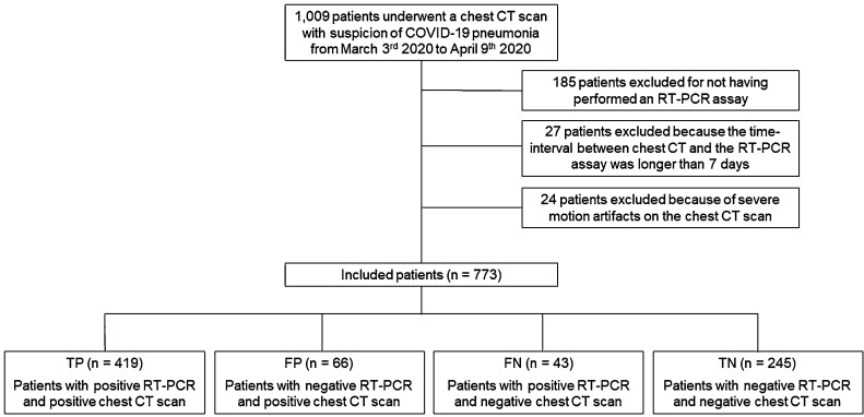
Methods: From March 4th to April 9th 2020, during the peak of the Italian COVID-19 epidemic, we enrolled a series of 773 patients that performed both non-contrast chest CT and RT-PCR with a time interval no longer than a week due to suspected SARS-CoV-2 infection. The diagnostic performance of CT was evaluated according to sensitivity, specificity, positive predictive value (PPV), negative predictive value (NPV) and diagnostic accuracy, considering RT-PCR as the reference standard. An analysis on the patients with discrepant CT scan and RT-PCR result and on the patient with both negative tests was performed.

Results: RT-PCR testing showed an overall positive rate of 59.8 %. CT sensitivity, specificity, PPV, NPV, and accuracy for SARS-CoV-2 infection were 90.7 % [95 % IC, 87.7%-93.2%], 78.8 % [95 % IC, 73.8-83.2%], 86.4 % [95 % IC, 76.1 %-88.9 %], 85.1 % [95 % IC, 81.0 %-88.4] and 85.9 % [95 % IC 83.2-88.3%], respectively. Twenty-five/66 (37.6 %) patients with positive CT and negative RT-PCR results and 12/245 (4.9 %) patients with both negative tests were nevertheless judged as positive cases by the clinicians based on clinical and epidemiological criteria and consequently treated.

Conclusions: In our experience, in a context of high pre-test probability, CT scan shows good sensitivity and a consistently higher specificity for the diagnosis of COVID-19 pneumonia than what reported by previous studies, especially when clinical and epidemiological features are taken into account.

Keywords: COVID-19; Diagnostic X-ray radiology; Sars-CoV-2; Tomography; X-ray computed.


Conflict of interest statement

The authors of this manuscript declare no relationships with any companies, whose products or services may be related to the subject matter of the article.

Figures

Fig. 1
Fig. 1
Flowchart of the study. Abbreviation: RT-PCR, reversal transcription polymerase chain reaction.
Fig. 2
Fig. 2
Chest CT images of the various pattern of COVID-19 pneumonia in true positive (TP) patients. A) A 36-year-old man with bilateral, peripheral patchy areas of ground-glass opacity (GGO) (axial view). B) A 42-year-old man with bilateral diffuse areas of GGO (axial view). C) A 45-year-old woman with bilateral diffuse areas of consolidation associated with GGO (axial view).
Fig. 3
Fig. 3
Chest CT features consistent with COVID-19 pneumonia in TP patients. A) A 60-year-old woman with bilateral patchy areas of GGO (coronal view). B) A 43-year-old woman with bilateral diffuse areas of consolidation associated with GGO (coronal view). C) A 63-year-old man with bilateral diffuse areas of crazy paving (axial view). D) A 39-year-old man with reversed halo sign in the left inferior lobe (axial view).
Fig. 4
Fig. 4
Chest CT features non consistent with COVID-19 pneumonia in true negative (TN) patients. Column A) A 32-year-old man with no parenchymal or mediastinal alteration (axial and coronal view). Column B) A 55-year-old woman with “tree-in-bud” alterations in the right upper lobe (axial and coronal view). Column C) A 85-year-old woman with bilateral pleural effusion and CT features consistent with congestive heart failure (CHF) (axial and coronal view).
Fig. 5
Fig. 5
Chest CT images of false positive (FP) patients. Column A) A 79-year-old woman hospitalized but not treated for COVID-19; an alternative diagnosis of bacterial pneumonia by E. Coli was formulated. Chest CT shows diffuse areas of consolidation associated with GGO with a prevalent perihilar distribution (axial and coronal view). Column B) A 57-year-old man hospitalized and treated for COVID-19. Chest CT shows bilateral, peripheral patchy areas of GGO with initial consolidation (axial and coronal view).
Fig. 6
Fig. 6
Chest CT images of FN patients with confounding features. Column A) A 29-year-old man with no parenchymal or mediastinal alteration (axial and coronal view). Column B) A 86-year-old with diffuse emphysema (axial and coronal view). Column C) A 75-year-old woman with bilateral pleural effusion, multiple areas of consolidation and CT features consistent with CHF (axial and coronal view).

References

    1. Coronaviridae Study Group of the International Committee on Taxonomy of Viruses The species severe acute respiratory syndrome-related coronavirus: classifying 2019-nCoV and naming it SARS-CoV-2. Nat. Microbiol. 2020;5(4):536–544. doi: 10.1038/s41564-020-0695-z. - DOI - PMC - PubMed
    1. Vermeiren C., Marchand-Senécal X., Sheldrake E. Comparison of Copan Eswab and FLOQswab for COVID-19 PCR diagnosis: working around a supply shortage. J. Clin. Microbiol. 2020;58(6) doi: 10.1128/JCM.00669-20. e00669-20. Published 2020 May 26. - DOI - PMC - PubMed
    1. Chung M., Bernheim A., Mei X. CT imaging features of 2019 novel coronavirus (2019-nCoV) Radiology. 2020;295(1):202–207. doi: 10.1148/radiol.2020200230. - DOI - PMC - PubMed
    1. Pan Y., Guan H. Imaging changes in patients with 2019-nCov. Eur. Radiol. 2020:1–2. doi: 10.1007/s00330-020-06713-z. [published online ahead of print, 2020 Feb 6] - DOI - PMC - PubMed
    1. Corman V.M., Landt O., Kaiser M. Detection of 2019 novel coronavirus (2019-nCoV) by real-time RT-PCR. Euro Surveill. 2020;25(3):2000045. doi: 10.2807/1560-7917.ES.2020.25.3.2000045. - DOI - PMC - PubMed

Publication types

MeSH terms