

doi: 10.1007/s00415-020-10103-2.
Epub 2020 Aug 1.
Causality in COVID-19-associated stroke: a uniform case definition for use in clinical research
Affiliations
- PMID: 32740765
- PMCID: PMC7395574
- DOI: 10.1007/s00415-020-10103-2
Item in Clipboard
Causality in COVID-19-associated stroke: a uniform case definition for use in clinical research
J Neurol.
2021 Mar.
No abstract available
Keywords: COVID-19; Cerebrovascular disease; Hemorrhagic stroke; Ischemic stroke; Neurological complications; SARS-CoV-2.
Conflict of interest statement
None reported.
Figures
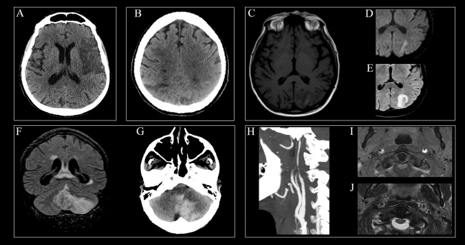
Neuroimaging features of COVID-19-associated stroke. a A 70-year-old man with critical COVID-19-related ARDS developed acute right-sided weakness. Brain CT showed a large fronto-insular ischemic lesion within the vascular territory of the left middle cerebral artery. b A 64-year-old man with COVID-19 infection developed multi-organ failure. Brain CT showed multiple recent ischemic lesions involving cortical-subcortical regions of both parietal lobes and centrum semiovale. c–e A 67-year-old man with COVID-19 and critical ARDS presented myocardial infarction 2 days after hospitalization. On day 14, he developed a tetraparesis. Brain and spine MRI were requested. As an incidental finding (given the final diagnosis of critical illness neuropathy), brain MRI showed a left parieto-occipital infarction, hyperintense on T1-weighted images (c) and bright on DWI (d) along the cortex, with subcortical white matter perilesional edema on FLAIR sequence (e). f–g A 72-year-old man diagnosed with COVID-19 presented with ataxia and vomiting. MRI showed infarction of the postero-inferior part of the left cerebellar hemisphere and the inferior part of the vermis in the territory of PICA (f, coronal FLAIR). Areas of hypointensity within the vermis, corresponding to blood degradation products, were also noted. The lesion underwent extensive hemorrhagic transformation with large parenchymal hematoma (g, axial CT). h–j A 58-year-old man with moderate COVID-19 presented with intense headache and neck pain. h CT angiography showed a long stenosis of the distal part of internal carotid artery bilaterally. MRI axial T1-weighted images obtained with fat saturation (i) and T2-weighted images (j) showed a narrowed eccentric flow void surrounded by a crescent-shaped subacute mural hematoma. This case of bilateral carotid dissection was previously reported by our group [12]. ARDS acute respiratory distress syndrome, CT computed tomography, DWI diffusion-weighted imaging, FLAIR fluid-attenuated inversion recovery, MRI magnetic resonance imaging, PICA posterior inferior cerebellar artery
References
Publication types
MeSH terms
LinkOut - more resources
Full Text Sources
Medical
Miscellaneous
