. 2021 May;73(5):1747-1763.
doi: 10.1002/hep.31486.

Ten-Eleven Translocation 1 Promotes Malignant Progression of Cholangiocarcinoma With Wild-Type Isocitrate Dehydrogenase 1

Affiliations

Ten-Eleven Translocation 1 Promotes Malignant Progression of Cholangiocarcinoma With Wild-Type Isocitrate Dehydrogenase 1

Xuewei Bai et al. Hepatology. 2021 May.

Abstract

Background and aims: Cholangiocarcinoma (CCA) is a highly lethal disease without effective therapeutic approaches. The whole-genome sequencing data indicate that about 20% of patients with CCA have isocitrate dehydrogenase 1 (IDH1) mutations, which have been suggested to target 2-oxoglutarate (OG)-dependent dioxygenases in promoting CCA carcinogenesis. However, the clinical study indicates that patients with CCA and mutant IDH1 have better prognosis than those with wild-type IDH1, further complicating the roles of 2-OG-dependent enzymes.

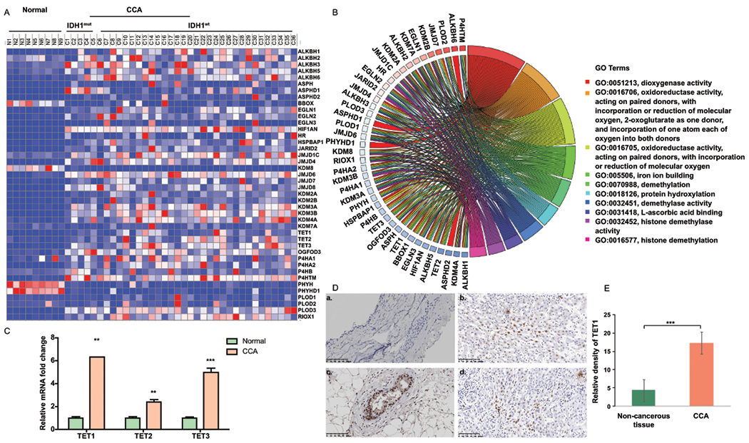
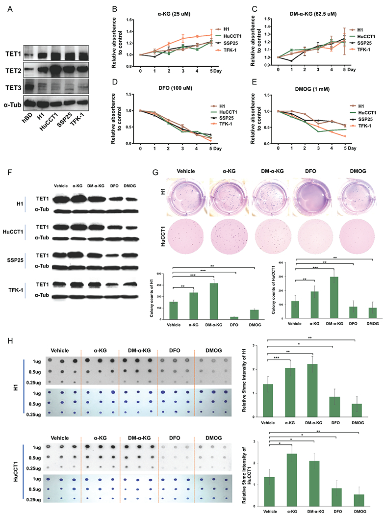
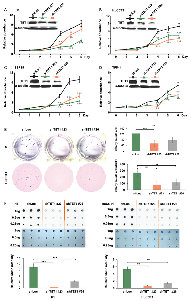
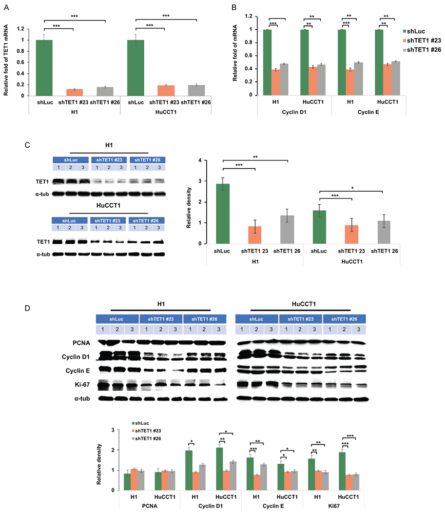
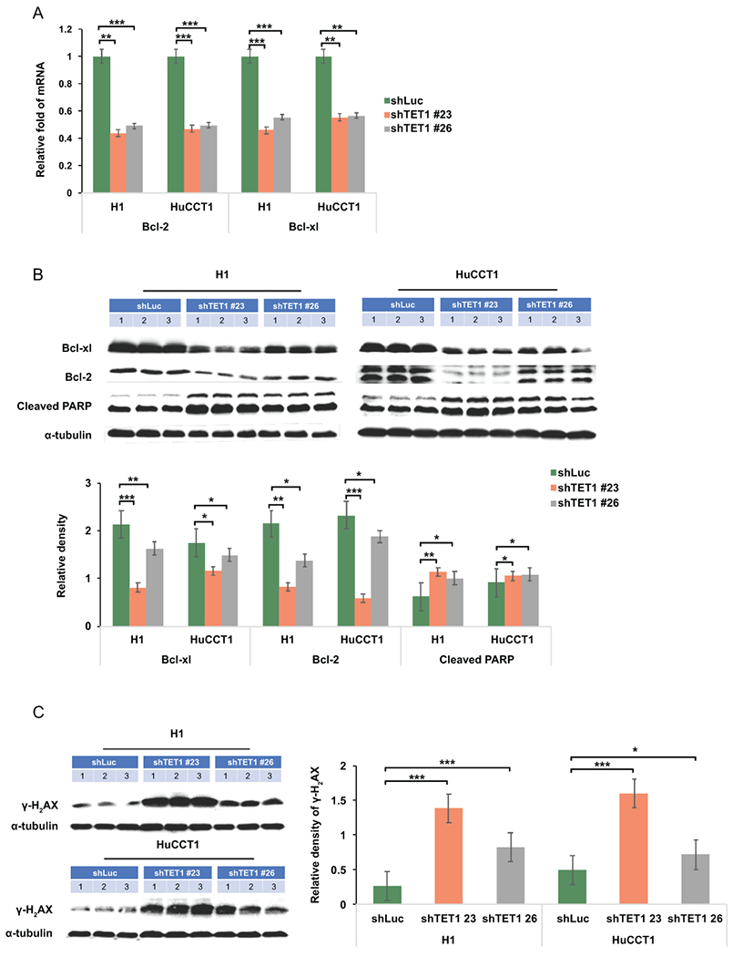
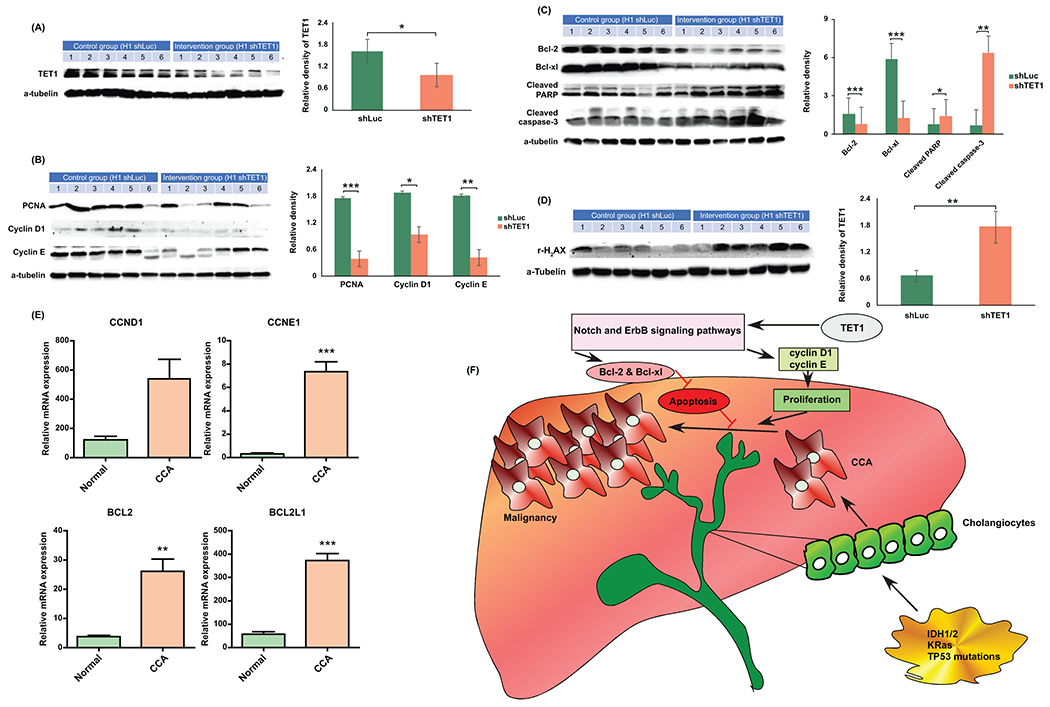
Approach and results: This study aimed to clarify if ten-eleven translocation 1 (TET1), which is one of the 2-OG-dependent enzymes functioning in regulating 5-hydroxymethylcytosine (5hmC) formation, is involved in CCA progression. By analyzing The Cancer Genome Atlas (TCGA) data set, TET1 mRNA was found to be substantially up-regulated in patients with CCA when compared with noncancerous bile ducts. Additionally, TET1 protein expression was significantly elevated in human CCA tumors. CCA cells were challenged with α-ketoglutarate (α-KG) and dimethyl-α-KG (DM-α-KG), which are cosubstrates for TET1 dioxygenase. The treatments with α-KG and DM-α-KG promoted 5hmC formation and malignancy of CCA cells. Molecular and pharmacological approaches were used to inhibit TET1 activity, and these treatments substantially suppressed 5hmC and CCA carcinogenesis. Mechanistically, it was found that knockdown of TET1 may suppress CCA progression by targeting cell growth and apoptosis through epigenetic regulation. Consistently, targeting TET1 significantly inhibited CCA malignant progression in a liver orthotopic xenograft model by targeting cell growth and apoptosis.

Conclusions: Our data suggest that expression of TET1 is highly associated with CCA carcinogenesis. It will be important to evaluate TET1 expression in CCA tumors before application of the IDH1 mutation inhibitor because the inhibitor suppresses 2-hydroxyglutarate expression, which may result in activation of TET, potentially leading to CCA malignancy.


Figures

Figure 1.
Figure 1.
TET1 expression is elevated in CCA patients. (A) Heat map for the expression of 2-oxogluratate dependent enzymes was generated by using TCGA data which includes 9 normal samples, 6 CCA tumors with IDH1 mutation, and 30 CCA tumors with IDH1 wild-type. (B) The association of molecular signaling pathways and functions with α-KG dependent enzymes. (C) Relative TET1, TET2, and TET3 mRNA expression levels were analyzed in human normal bile ducts (n=9) and CCA patients (n=36) by using TCGA data. (D) The representative TET1 immunohistochemistry (IHC) images were shown in non-cancerous bile duct (a) and CCA samples(b-d). (E) Semi-quantification of TET1 IHC staining data was calculated by using the Image J software. **, p<0.01; ***, p<0.001 when compared with relevant control.
Figure 2.
Figure 2.
CCA cell growth was modulated by TET1 activity. (A) The expression levels of TETs in normal cholangiocytes (hBD) and CCA cell lines (H1, HuCCT1, SSP25 and TFK-1) were examined by western blotting. The results of cell growth rates were analyzed in CCA cells treated with (B) α-KG, (C) DM-α-KG, (D) DFO, and (E) DMOG as indicated. The medium including agents were changed every other day. (F) TET1 protein expression was determined in CCA cells treated with α-KG, DM-α-KG, DFO and DMOG for 3 days. α-Tubulin served as loading control. (G)The colony formation abilities were evaluated in H1 and HuCCT1 in the presence or absence of α-KG, DM-α-KG, DFO and DMOG. Graph results indicate the average of triplicate experiments. (H) Dot blot assays were adopted to analyze 5hmC (black spots) levels in H1 (upper panel) and HuCCT1 (lower panel) cells treated as indicated for 3 days. The methylene blue staining data (blue spots) served as DNA loading control. Graph next to the 5hmC image is quantification of 5hmC. *p<0.05; **p<0.01; ***p<0.001.
Figure 3.
Figure 3.
Targeting TET1 specifically with 2 shRNAs reduced CCA cell growth. (A-D) Relative cell growth rates were analyzed (A) H1, (B) HuCCT1, (C) SSP25 and (D) TFK-1 treated with shRNA-luciferase (shLuc), shRNA-TET1#23 (shTET1#23), and shRNA-TET1#26 (shTET1#26) by MTT assay. The relative absorbance is the ratio of the absolute value of absorbance at the specific time point to the day 0. The western blotting data was used to verify TET1 down-regulation. (E) Knockdown of TET1 reduced soft-agar colony formation abilities of H1 and HuCCT1. (F) Dot blot results of 5hmC in H1 and HuCCT1 treated with shLuc, shTET1#23, and shTET1#26. Methylene blue staining data served as DNA loading control. Bottom graph results show the quantification of 5hmC. *p<0.05; **p<0.01; ***p<0.001.
Figure 4.
Figure 4.
Knockdown of TET1 inhibited cell proliferation associated signaling pathways. (A) TET1, (B) cyclin D1, and cyclin E1 mRNA expression levels were examined in shLuc-, shTET1#23-, and shTET1#26-treated H1 and HuCCT1 cells. (C) TET1 protein levels were verified in H1 and HuCCT1 cells as indicated. (D) PCNA, cyclin D1, cyclin E1, and a-tubulin were determined in control and TET1-knockdown H1 and HuCCT1 CCA cells. Graph results indicate the average of triplicate semi-quantifications of western blotting data. *p<0.05; **p<0.01; ***p<0.001.
Figure 5.
Figure 5.
TET1 down-regulation suppressed anti-apoptotic protein expression but had no impacts on pro-apoptotic genes. (A) The mRNA expression of anti-apoptosis protein Bcl-2 and Bcl-XL was examined in CCA cells treated as indicated. (B) BCL-XL, Bcl2, cleaved PARP, and (C) γ-H2aX were determined in shLuc-, shTET1#23-, and shTET1#26-treated H1 and HuCCT1 cells. α-tubulin served as loading control. Graph data shows semi-quantification of western blotting results. *p<0.05; **p<0.01; ***p<0.001.
Figure 6.
Figure 6.
Knockdown of TET1 repressed CCA progression in vivo. (A) Gross images of xenograft tumors derived from shLuc- and shTET1#23-treated H1 CCA cells. (B) The representative H&E staining images (400× and 1000×) in shLuc and shTET1#23 groups. (C) The tumor weight and ratio of tumor weight to body weight are significantly decreased in shTET#23 group when compared with control one, n=6.
Figure 7.
Figure 7.
Targeting TET1 inhibited cell growth and induced cell apoptosis in vivo. (A) TET1 expression was verified in shLuc and shTET1#23 H1 orthotopic xenograft tumors. (B) Cell growth associated proteins including PCNA, Cyclin D1 and Cyclin E were analyzed in shLuc- and shTET1#23 H1 xenograft tumors. (C) Anti-apoptotic proteins, Bcl2 and Bcl-XL were decreased in shTET1#23-treated tumors. Cleaved caspase 3 and PARP were elevated in TET1 knockdown tumors. (D) The DNA damage marker, γ-H2aX was increased in TET1 kncokdown tumors as well. Graph data shows the semi-quantification results of relevant proteins. (E) The mRNA expression of cyclin D1 (CCND1), cyclin E1 (CCNE1), Bcl-2 (BCL2), and Bcl-XL (BCL2L1) was analyzed in human normal (n=9) and CCA tissues (n=36) by using TCGA data. (F) A summary cartoon illustrating how TET1 may modulate CCA progression. *p<0.05; **p<0.01; ***p<0.001.

References

    1. Razumilava N, Gores GJ. Cholangiocarcinoma. Lancet 2014;383:2168–2179. - PMC - PubMed
    1. Jiao Y, Pawlik TM, Anders RA, Selaru FM, Streppel MM, Lucas DJ, Niknafs N, et al. Exome sequencing identifies frequent inactivating mutations in BAP1, ARID1A and PBRM1 in intrahepatic cholangiocarcinomas. Nat Genet 2013;45:1470–1473. - PMC - PubMed
    1. Chan-On W, Nairismagi ML, Ong CK, Lim WK, Dima S, Pairojkul C, Lim KH, et al. Exome sequencing identifies distinct mutational patterns in liver fluke-related and non-infection-related bile duct cancers. Nat Genet 2013;45:1474–1478. - PubMed
    1. Saha SK, Parachoniak CA, Ghanta KS, Fitamant J, Ross KN, Najem MS, Gurumurthy S, et al. Mutant IDH inhibits HNF-4alpha to block hepatocyte differentiation and promote biliary cancer. Nature 2014;513:110–114. - PMC - PubMed
    1. Dang L, White DW, Gross S, Bennett BD, Bittinger MA, Driggers EM, Fantin VR, et al. Cancer-associated IDH1 mutations produce 2-hydroxyglutarate. Nature 2009;462:739–744. - PMC - PubMed

Publication types

MeSH terms