Review
. 2020 Aug 2;15(1):11.
doi: 10.1186/s13064-020-00147-5.

Synapse development and maturation at the drosophila neuromuscular junction

Affiliations
Review

Synapse development and maturation at the drosophila neuromuscular junction

Vivian T Chou et al. Neural Dev. .

Abstract

Synapses are the sites of neuron-to-neuron communication and form the basis of the neural circuits that underlie all animal cognition and behavior. Chemical synapses are specialized asymmetric junctions between a presynaptic neuron and a postsynaptic target that form through a series of diverse cellular and subcellular events under the control of complex signaling networks. Once established, the synapse facilitates neurotransmission by mediating the organization and fusion of synaptic vesicles and must also retain the ability to undergo plastic changes. In recent years, synaptic genes have been implicated in a wide array of neurodevelopmental disorders; the individual and societal burdens imposed by these disorders, as well as the lack of effective therapies, motivates continued work on fundamental synapse biology. The properties and functions of the nervous system are remarkably conserved across animal phyla, and many insights into the synapses of the vertebrate central nervous system have been derived from studies of invertebrate models. A prominent model synapse is the Drosophila melanogaster larval neuromuscular junction, which bears striking similarities to the glutamatergic synapses of the vertebrate brain and spine; further advantages include the simplicity and experimental versatility of the fly, as well as its century-long history as a model organism. Here, we survey findings on the major events in synaptogenesis, including target specification, morphogenesis, and the assembly and maturation of synaptic specializations, with a emphasis on work conducted at the Drosophila neuromuscular junction.

Keywords: Bouton addition; Cell-adhesion molecules; Drosophila melanogaster; Neuromuscular junction; Presynaptic active zone; Synapse; Synaptic plasticity; Trans-synaptic signaling.


Conflict of interest statement

The authors declare that they have no competing interests.

Figures

Fig. 1
Fig. 1
Organization of the pre- and postsynaptic cytomatrix. a Drawing of the larval musculature, cartoons of type I NMJ/boutons and immunostaining of NMJ branches in muscle 6/7 marked with α-Brp (green) for active zones and α-HRP (magenta) for overall shape. b (Top) Electron micrograph displaying presynaptic AZ components, including clustered SVs and the electron-dense Brp T-bar, as well as the membrane folds of the SSR, which is thought to be structurally and functionally analogous to mammalian dendritic spines. The expected localization of iGluRs is also indicated. (Bottom) Schematic of selected pre- and postsynaptic components. Communication between compartments is mediated by trans-synaptic interactions, such as the Dnrx1-Dnlg1 complex, through which the presynaptic AZ protein Syd-1 acts to regulate the maturation of iGluR receptors. GluR clustering is also mediated by dPak via Pix
Fig. 2
Fig. 2
Overview of bouton growth and synaptic maturation. Addition of boutons is initiated by membrane outgrowth (a), followed by size expansion (b). (Top) Within minutes, AZ precursors are formed as early components such as Syd-1 and Liprin-α accumulate. (Bottom) Immunostaining of type I NMJ marked with α-Dlg for post synaptic SSR and α-HRP for presynaptic area. Immature boutons lacking postsynaptic structure (ghost boutons) are indicated (Ci). Maturation occurs as remaining components of the pre- and postsynaptic specializations are recruited, including GluRIIA-type receptors (Cii); Brp, Cac, and SVs (Ciii); and GluRIIB-type receptors (Civ). Inset (c′) shows bidirectional trans-synaptic pathways that are known to be important for synaptic development. Major pathways include both canonical and non-canonical Wnt and BMP signaling, while pathways mediated by Syt4, LAR, FMR1, Jeb, and MTG have also been described. The FGF pathway has also been reported, but pathways details, e.g. directionality, have not been defined
Fig. 3
Fig. 3
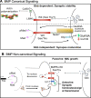
BMP Canonical and Non-Canonical signaling at the NMJ. a BMP canonical signaling includes a paracrine, retrograde mechanism whereby muscle-derived Gbb is released across the synapse where it activates BMPRs Wit and Sax/Tkv triggering internalization. Internalized Gbb is trafficked to the neuronal nucleus where it activates phosphorylation of the Mad to pMad to activate transcription of Trio-GEF promoting overall NMJ growth. A second autocrine canonical pathway involves release of Cmpy bound Gbb from the neuron where it sequestered by α2δ-3, a calcium channel subunit. Neuronally derived Gbb then binds BMPRs and causes transcriptional changes similar to the paracrine canonical pathway to regulate synaptic homeostasis and signal transmission. b BMP non-canonical signaling pathways include those that are Gbb dependent and independent. Gbb dependent non-canonical signaling involves retrograde release of Gbb where it is bound by Wit. LIMK acts as a Wit effector to inhibit Cofilin which normally promotes actin dynamics through filament severing. In a Gbb independent non-canonical mechanism, GluRIIA containing iGluRs form a positive feedback loop with local presynaptic pMad. This mechanism depends on Wit and Sax/Tkv, but not on transcription and is inhibited by Dlp (Glypican)

References

    1. Saleeba C, Dempsey B, Le S, Goodchild A, McMullan S. A Student’s guide to neural circuit tracing. Front Neurosci. 2019;13:1–19. - PMC - PubMed
    1. Herculano-Houzel S. The human brain in numbers: a linearly scaled-up primate brain. Front Hum Neurosci. 2009;3:31. - PMC - PubMed
    1. Colón-Ramos DA. Synapse formation in developing neural circuits. Curr Top Dev Biol. 2009;87(09):53–79. - PMC - PubMed
    1. Comer JD, Alvarez S, Butler SJ, Kaltschmidt JA. Commissural axon guidance in the developing spinal cord: from Cajal to the present day. Neural Dev. 2019;14(1):1–16. - PMC - PubMed
    1. Chia PH, Li P, Shen K. Cell biology in neuroscience: cellular and molecular mechanisms underlying presynapse formation. J Cell Biol. 2013;203(1):11–22. - PMC - PubMed

Publication types

MeSH terms

Substances

LinkOut - more resources