

Assessment of Small Pulmonary Blood Vessels in COVID-19 Patients Using HRCT
- PMID: 32741657
- PMCID: PMC7381940
- DOI: 10.1016/j.acra.2020.07.019
Assessment of Small Pulmonary Blood Vessels in COVID-19 Patients Using HRCT
Abstract
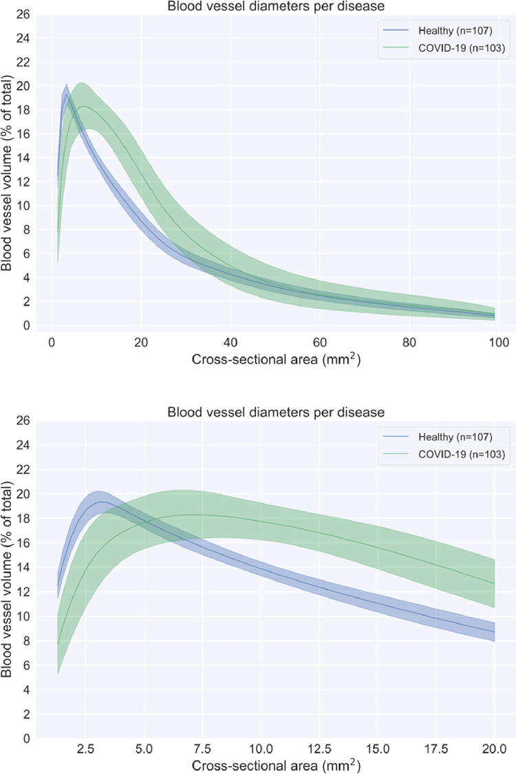
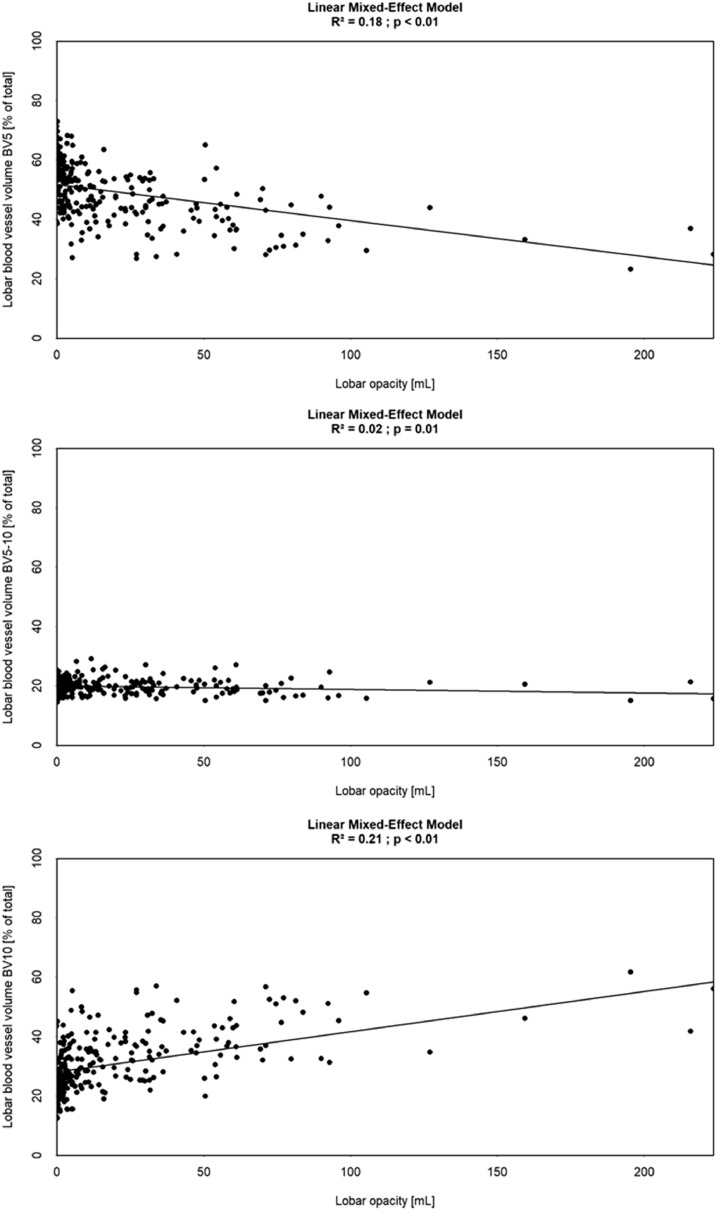
Rationale and objectives: Mounting evidence supports the role of pulmonary hemodynamic alternations in the pathogenesis of COVID-19. Previous studies have demonstrated that changes in pulmonary blood volumes measured on computed tomography (CT) are associated with histopathological markers of pulmonary vascular pruning, suggesting that quantitative CT analysis may eventually be useful in the assessment pulmonary vascular dysfunction more broadly.
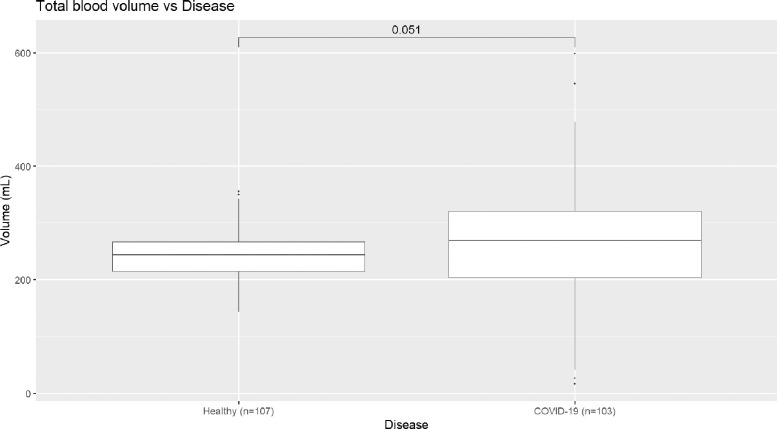
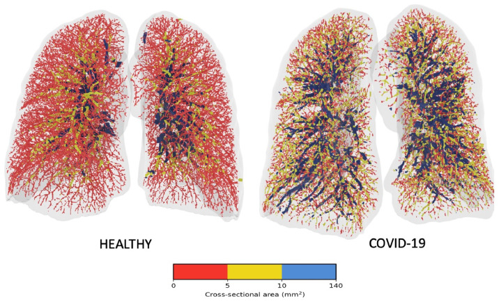
Materials and methods: Building upon previous work, automated quantitative CT measures of small blood vessel volume and pulmonary vascular density were developed. Scans from 103 COVID-19 patients and 107 healthy volunteers were analyzed and their results compared, with comparisons made both on lobar and global levels.
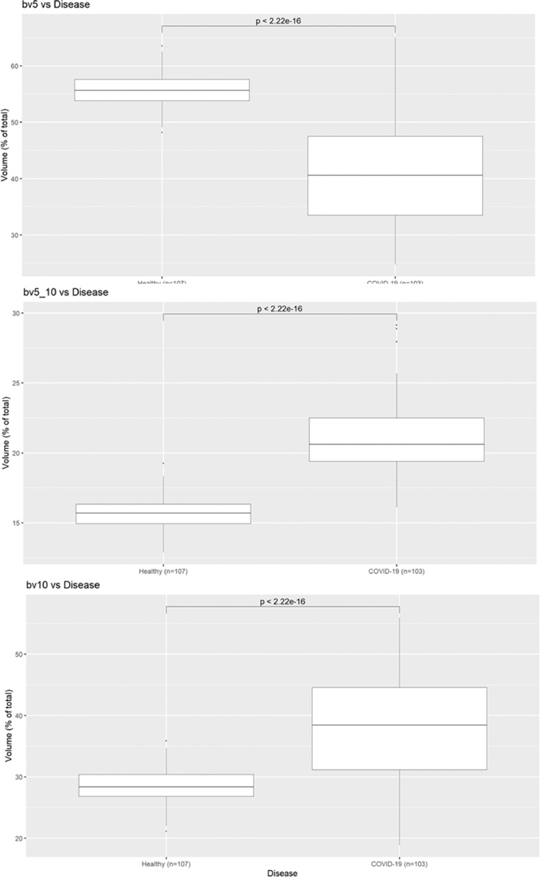
Results: Compared to healthy volunteers, COVID-19 patients showed significant reduction in BV5 (pulmonary blood volume contained in blood vessels of <5 mm2) expressed as BV5/(total pulmonary blood volume; p < 0.0001), and significant increases in BV5-10 and BV 10 (pulmonary blood volumes contained in vessels between 5 and 10 mm2 and above 10 mm2, respectively, p < 0.0001). These changes were consistent across lobes.
Conclusion: COVID-19 patients display striking anomalies in the distribution of blood volume within the pulmonary vascular tree, consistent with increased pulmonary vasculature resistance in the pulmonary vessels below the resolution of CT.
Keywords: COVID-19; Pulmonary vascular disease; Quantitative CT metrics.
Copyright © 2020 The Association of University Radiologists. Published by Elsevier Inc. All rights reserved.
Figures
References
Publication types
MeSH terms
LinkOut - more resources
Full Text Sources
Other Literature Sources
