. 2020 Dec;22(12):1956-1966.
doi: 10.1038/s41436-020-0907-1. Epub 2020 Aug 3.

Genetic dissection of spermatogenic arrest through exome analysis: clinical implications for the management of azoospermic men

Affiliations

Genetic dissection of spermatogenic arrest through exome analysis: clinical implications for the management of azoospermic men

Csilla Krausz et al. Genet Med. 2020 Dec.

Abstract

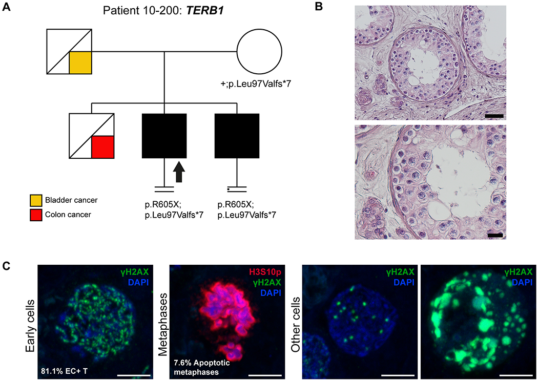
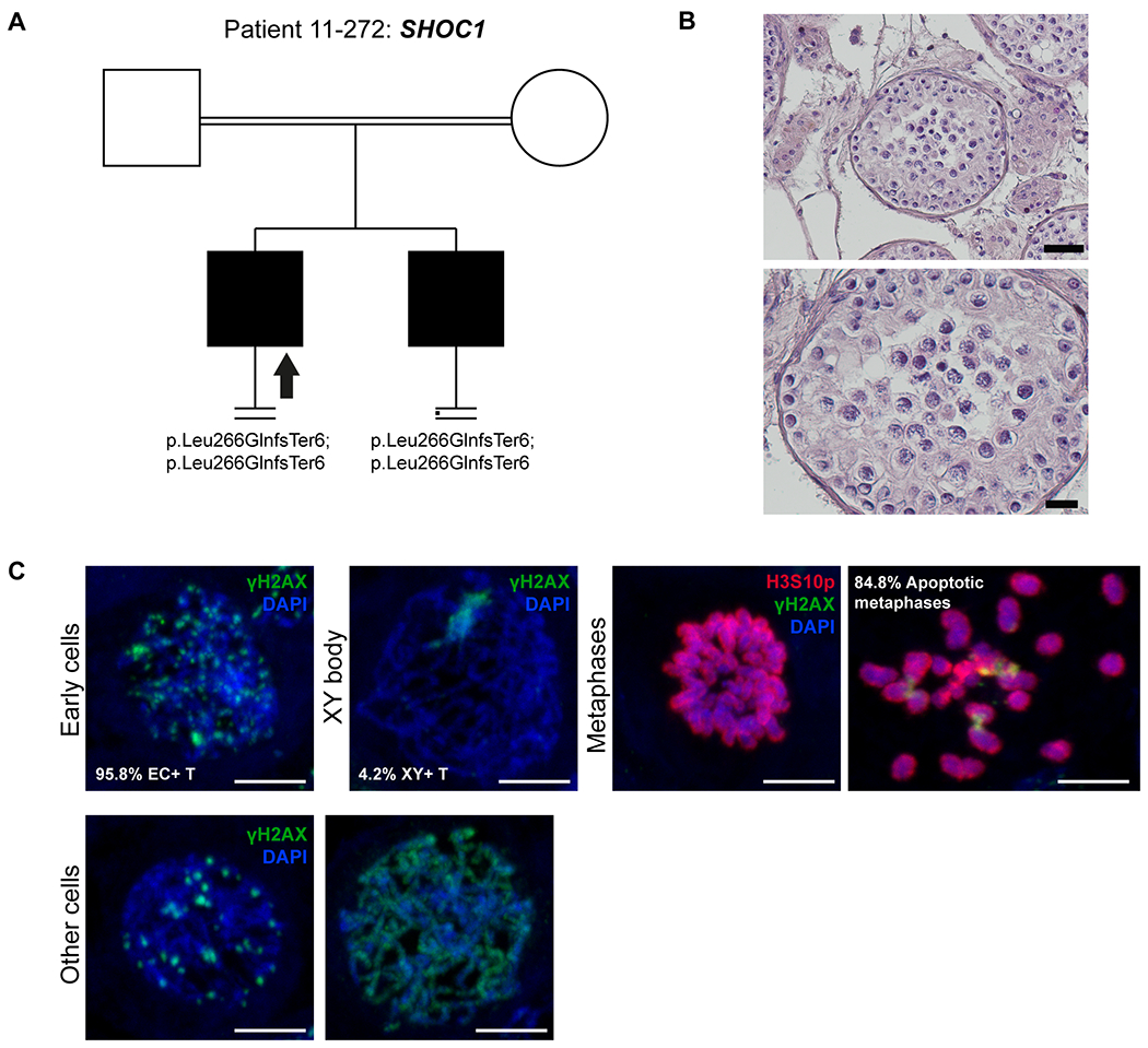
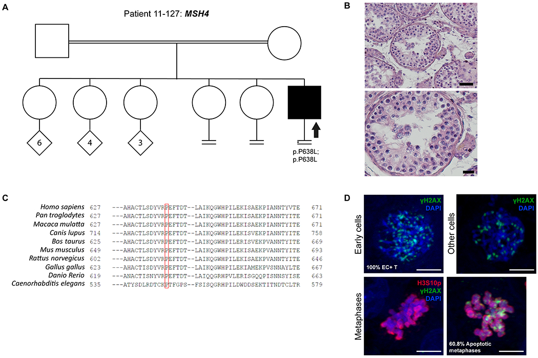
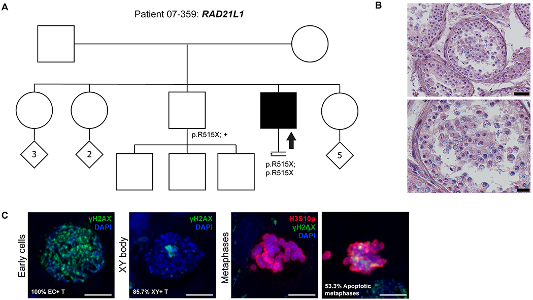
Purpose: Azoospermia affects 1% of men and it can be the consequence of spermatogenic maturation arrest (MA). Although the etiology of MA is likely to be of genetic origin, only 13 genes have been reported as recurrent potential causes of MA.

Methods: Exome sequencing in 147 selected MA patients (discovery cohort and two validation cohorts).

Results: We found strong evidence for five novel genes likely responsible for MA (ADAD2, TERB1, SHOC1, MSH4, and RAD21L1), for which mouse knockout (KO) models are concordant with the human phenotype. Four of them were validated in the two independent MA cohorts. In addition, nine patients carried pathogenic variants in seven previously reported genes-TEX14, DMRT1, TEX11, SYCE1, MEIOB, MEI1, and STAG3-allowing to upgrade the clinical significance of these genes for diagnostic purposes. Our meiotic studies provide novel insight into the functional consequences of the variants, supporting their pathogenic role.

Conclusion: Our findings contribute substantially to the development of a pre-testicular sperm extraction (TESE) prognostic gene panel. If properly validated, the genetic diagnosis of complete MA prior to surgical interventions is clinically relevant. Wider implications include the understanding of potential genetic links between nonobstructive azoospermia (NOA) and cancer predisposition, and between NOA and premature ovarian failure.

Keywords: azoospermia; genetics; male infertility; meiosis; spermatogenesis.


Conflict of interest statement

COMPETING OF INTEREST

The authors declare no competing interests

Figures

Figure 1.
Figure 1.. Investigation of patient 10-200 carrying the TERB1 variant.
A) The pedigree structure shows the segregation of the TERB1 variants (p.R605Ter and p.Leu97Valfs*7). B) H&E staining of histological sections from the testis biopsy of the patient carrying the TERB1 variant. Scale bar on the upper image represents 50 μm and on the lower image 20 μm. C) Immunofluorescent staining of histological sections from the testis biopsy of the patient carrying the TERB1 variant using γH2AX (Green, marker for DSBs and XY body), Histone 3 Serine 10 phosphorylation (H3S10p, red, marker of M-phase), and DAPI (blue). Scale bar represents 5μm. In this patient only a few metaphases were observed (1.1 metaphases/mm2), which were below the normal threshold, based on samples containing meiotic metaphases (4.0-11.5 metaphases/mm2), thus representing the mitotic metaphases of spermatogonia. Early cells were observed in almost all the counted tubules. No XY bodies were detected, indicating that the cells fail to reach the pachytene stage. Most of the early cells displayed an aberrant pattern of γH2AX spots. Some cells displayed an aberrant pattern whereby γH2AX patches covered the entire nucleus.
Figure 2.
Figure 2.. Investigation of patient 11-272 carrying the SHOC1 variant.
A) The pedigree structure shows the segregation of the SHOC1 variant (p.Leu266GlnfsTer6). B) H&E staining of histological sections from the testis biopsy of the patient carrying the SHOC1 variant. Scale bar on the upper image represents 50 μm and on the lower image 20 μm. C) Immunofluorescent staining of histological sections from the testis biopsy of the patient’s brother carrying the SHOC1 variant using γH2AX (Green), H3S10p (red), and DAPI (blue). Scale bar represents 5μm. Early cells were observed in 95.8% of the round tubules that were counted. Extremely reduced XY body positive tubules were observed, indicating that the cells rarely reach the pachytene stage. A high density of metaphases was observed (29 metaphases/mm2) with a high percentage of apoptotic metaphases (84.8%). The organization of the apoptotic metaphases appeared to be more chaotic, as the chromosomes within these metaphases were dispersed. The early cells displayed an aberrant pattern of γH2AX spots. Some cells displayed an aberrant pattern whereby γH2AX is localized on the entire axis of the DNA strands.
Figure 3.
Figure 3.. Investigation of patient 11-127 carrying the MSH4 variant.
A) The pedigree structure shows the segregation of the MSH4 variant (p.P638L). B) H&E staining of histological sections from the testis biopsy of the patient carrying the MSH4 variant. Scale bar on the upper image represents 50 μm and on the lower image 20 μm. C) Sequence alignment of MSH4 protein orthologs, performed by HomoloGene. Red box highlights the Proline that is changed to Leucine in the MSH4 variant, which is highly conserved among species. D) Immunofluorescent staining of histological sections from the testis biopsy of the patient carrying the MSH4 variant using γH2AX (Green), H3S10p (red), and DAPI (blue). Scale bar represents 5μm. Early cells were observed in 100% of the round tubules that were counted. In a striking number of early cells we observed a spotty γH2AX pattern. No XY bodies were observed in the tubules that were counted for this patient. The metaphase density is higher (15.2 metaphases/mm2) than the previously described control group. This indicates that there are meiotic metaphases present in this patient despite the lack of XY bodies. In addition, 60.8 % of the metaphases were positive for γH2AX.
Figure 4.
Figure 4.. Investigation of patient 07-359 carrying the RAD21L1 variant.
A) The pedigree structure shows the segregation of the RAD21L1 variant (p.R515X). B) H&E staining of histological sections from the testis biopsy of the patient carrying the RAD21L1 variant. Scale bar on the upper image represents 50 μm and on the lower image 20 μm. C) Immunofluorescent staining of histological sections from the testis biopsy of the patient carrying the SHOC1 variant using γH2AX (Green), H3S10p (red), and DAPI (blue). Scale bar represents 5μm. Early cells were observed in 100% of the round tubules that were counted. XY bodies were present in 85.5% of the tubules. The metaphase density is comparable to the control group (9.4 metaphases/mm2) however, a high percentage of these metaphases are apoptotic (53.3%).

References

    1. Krausz C, Riera-Escamilla A. Genetics of male infertility. Nat Rev Urol. 2018;15(6):369–384. doi:10.1038/s41585-018-0003-3. - DOI - PubMed
    1. Tournaye H, Krausz C, Oates RD. Novel concepts in the aetiology of male reproductive impairment. lancet Diabetes Endocrinol July 2016. doi:10.1016/S2213-8587(16)30040-7. - DOI - PubMed
    1. Oud MS, Volozonoka L, Smits RM, Vissers LELM, Ramos L, Veltman JA. A systematic review and standardized clinical validity assessment of male infertility genes. Hum Reprod. 2019;34(5):932–941. doi:10.1093/humrep/dez022. - DOI - PMC - PubMed
    1. van der Bijl N, Ropke A, Biswas U, et al. Mutations in the stromal antigen 3 (STAG3) gene cause male infertility due to meiotic arrest. Hum Reprod. November 2019. doi:10.1093/humrep/dez204. - DOI - PubMed
    1. Riera-Escamilla A, Enguita-Marruedo A, Moreno-Mendoza D, et al. Sequencing of a “mouse azoospermia” gene panel in azoospermic men: identification of RNF212 and STAG3 mutations as novel genetic causes of meiotic arrest. Hum Reprod. 2019;34(6):978–988. doi:10.1093/humrep/dez042. - DOI - PubMed

Publication types