

Increased levels of miR-124 in human dental pulp stem cells alter the expression of neural markers
- PMID: 32742271
- PMCID: PMC7387844
- DOI: 10.1016/j.joto.2019.04.001
Increased levels of miR-124 in human dental pulp stem cells alter the expression of neural markers
Abstract
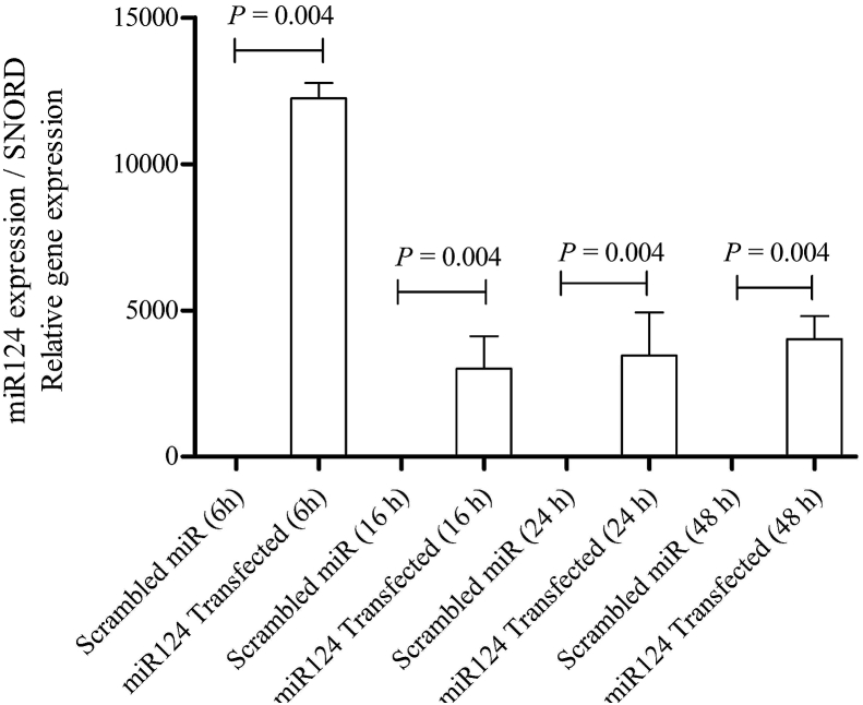
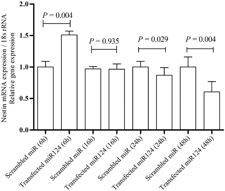
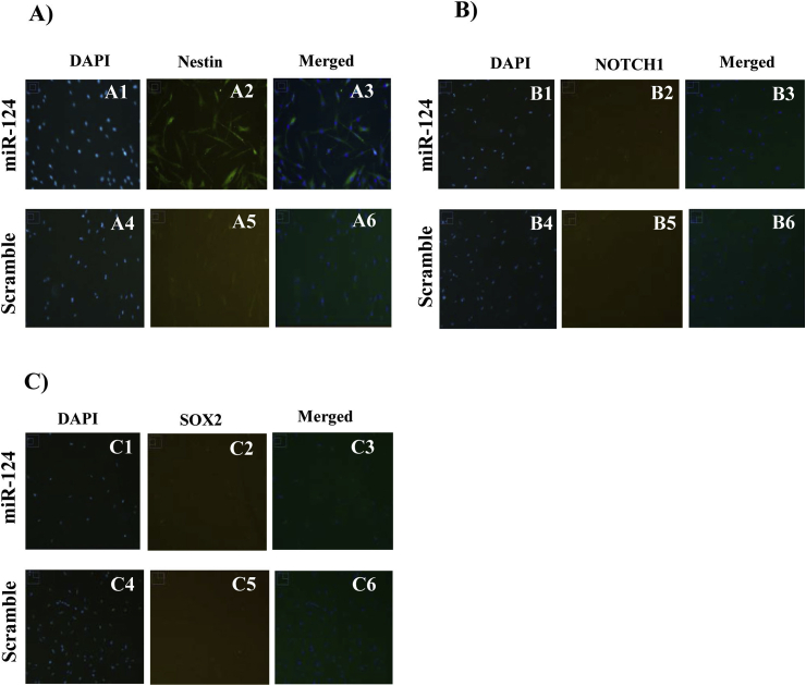
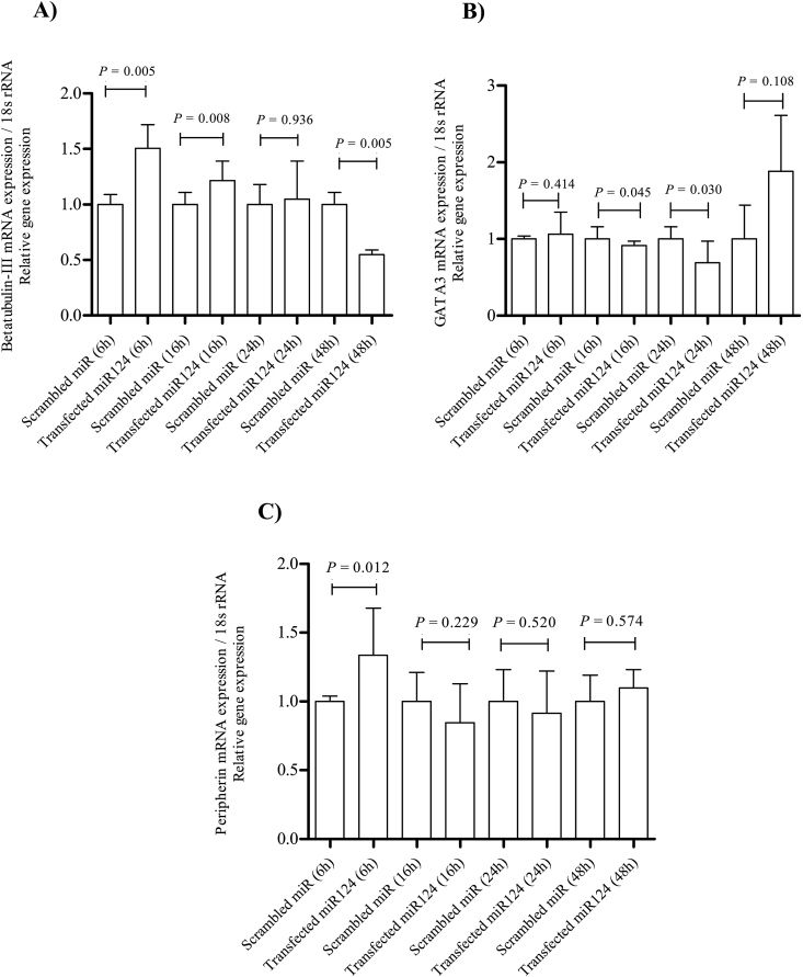
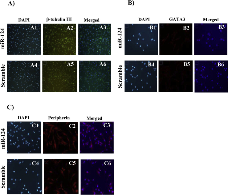
Auditory neuropathy is the particular form of deafness in humans which cannot be treated by replacement therapy. Human dental pulp stem cells (hDPSCs) are derived from an ectomesenchymal neural crest cell population. Therefore, they possess a promising capacity for neuronal differentiation and repair. miR-124, a key regulator of neuronal development in the inner ear, is expressed at high levels in auditory and vestibular neurons. Here, we evaluated the possible effect of miR-124 in alteration of neural protein markers expression. Using quantitative reverse transcription-PCR (qRT-PCR) analyses and immunofluorescence staining, we studied the expression patterns of neural progenitor markers (Nestin, NOTCH1, and SOX2) and neural markers (β-tubulin III, GATA-3, and peripherin) upon transfection of hDPSCs with miR-124. The qRT-PCR results showed that Nestin was upregulated 6 h post-transfection. In contrast, Nestin expression exhibited a decreasing trend 24 h and 48 h post-transfection. Higher levels of β-tubulin III, 6 h and 16 h post transfection in RNA level as compared with control cells, were determined in transfected DPSCs. However, β-tubulin-III expression decreased 48 h post-transfection. The immunoflourescence results indicated that transfection of hDPSCs with miR-124, only affected Nestin among the studied neural progenitor and neural marker expression in protein level.
Keywords: DPSCs; Nestin; Sensorineural hearing loss; Spiral ganglion neurons; basic fibroblast growth factor, bFGF; bone morphogenetic protein 4, BMP4; bovin serum albumin, BSA; brain derived neurotrophic factor, BDNF; epidermal growth factor, EGF; human dental pulp stem cells, hDPSCs; miR-124; neurotrophin-3, NT3; quantitative reverse transcription-PCR, qRT-PCR; sonic hedgehog, SHH; spiral ganglion neurons, SGNs.
© 2019 PLA General Hospital Department of Otolaryngology Head and Neck Surgery. Production and hosting by Elsevier (Singapore) Pte Ltd.
Figures
References
-
- Caccamo D.V., Herman M.M., Frankfurter A., Katsetos C.D., Collins V.P., Rubinstein L.J. An immunohistochemical study of neuropeptides and neuronal cytoskeletal proteins in the neuroepithelial component of a spontaneous murine ovarian teratoma. Primitive neuroepithelium displays immunoreactivity for neuropeptides and neuron-associated beta-tu. Am. J. Pathol. 1989;135(5):801–813. http://www.ncbi.nlm.nih.gov/pubmed/2817080 Retrieved from. - PMC - PubMed
-
- Chen W., Johnson S.L., Marcotti W., Andrews P.W., Moore H.D., Rivolta M.N. Human fetal auditory stem cells can be expanded in vitro and differentiate into functional auditory neurons and hair cell-like cells. Stem Cell. 2009;27(5):1196–1204. - PubMed
LinkOut - more resources
Full Text Sources
