High tibial osteotomy with human umbilical cord blood-derived mesenchymal stem cells implantation for knee cartilage regeneration
- PMID: 32742568
- PMCID: PMC7360989
- DOI: 10.4252/wjsc.v12.i6.514
High tibial osteotomy with human umbilical cord blood-derived mesenchymal stem cells implantation for knee cartilage regeneration
Abstract
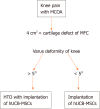
Background: High tibial osteotomy (HTO) is a well-established method for the treatment of medial compartment osteoarthritis of the knee with varus deformity. However, HTO alone cannot adequately repair the arthritic joint, necessitating cartilage regeneration therapy. Cartilage regeneration procedures with concomitant HTO are used to improve the clinical outcome in patients with varus deformity.
Aim: To evaluate cartilage regeneration after implantation of allogenic human umbilical cord blood-derived mesenchymal stem cells (hUCB-MSCs) with concomitant HTO.
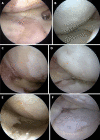
Methods: Data for patients who underwent implantation of hUCB-MSCs with concomitant HTO were evaluated. The patients included in this study were over 40 years old, had a varus deformity of more than 5°, and a full-thickness International Cartilage Repair Society (ICRS) grade IV articular cartilage lesion of more than 4 cm2 in the medial compartment of the knee. All patients underwent second-look arthroscopy during hardware removal. Cartilage regeneration was evaluated macroscopically using the ICRS grading system in second-look arthroscopy. We also assessed the effects of patient characteristics, such as trochlear lesions, age, and lesion size, using patient medical records.
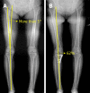
Results: A total of 125 patients were included in the study, with an average age of 58.3 ± 6.8 years (range: 43-74 years old); 95 (76%) were female and 30 (24%) were male. The average hip-knee-ankle (HKA) angle for measuring varus deformity was 7.6° ± 2.4° (range: 5.0-14.2°). In second-look arthroscopy, the status of medial femoral condyle (MFC) cartilage was as follows: 73 (58.4%) patients with ICRS grade I, 37 (29.6%) with ICRS grade II, and 15 (12%) with ICRS grade III. No patients were staged with ICRS grade IV. Additionally, the scores [except International Knee Documentation Committee (IKDC) at 1 year] of the ICRS grade I group improved more significantly than those of the ICRS grade II and III groups.
Conclusion: Implantation of hUCB-MSCs with concomitant HTO is an effective treatment for patients with medial compartment osteoarthritis and varus deformity. Regeneration of cartilage improves the clinical outcomes for the patients.
Keywords: Allogeneic; Arthroscopy; Cartilage regeneration; High tibial osteotomy; Human umbilical cord blood-derived mesenchymal stem cells; Osteoarthritic knees.
©The Author(s) 2020. Published by Baishideng Publishing Group Inc. All rights reserved.
Conflict of interest statement
Conflict-of-interest statement: We have no financial relationships to disclose.
Figures


References
-
- Shiomi T, Nishii T, Tanaka H, Yamazaki Y, Murase K, Myoui A, Yoshikawa H, Sugano N. Loading and knee alignment have significant influence on cartilage MRI T2 in porcine knee joints. Osteoarthritis Cartilage. 2010;18:902–908. - PubMed
-
- Cerejo R, Dunlop DD, Cahue S, Channin D, Song J, Sharma L. The influence of alignment on risk of knee osteoarthritis progression according to baseline stage of disease. Arthritis Rheum. 2002;46:2632–2636. - PubMed
-
- Glyn-Jones S, Palmer AJ, Agricola R, Price AJ, Vincent TL, Weinans H, Carr AJ. Osteoarthritis. Lancet. 2015;386:376–387. - PubMed
LinkOut - more resources
Full Text Sources

