

Established paths and new avenues: a review of the main radiological techniques for investigating sarcopenia
- PMID: 32742955
- PMCID: PMC7378089
- DOI: 10.21037/qims.2019.12.15
Established paths and new avenues: a review of the main radiological techniques for investigating sarcopenia
Abstract
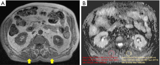
Sarcopenia is a clinical condition mainly affecting the elderly that can be associated in a long run with severe consequences like malnutrition and frailty. Considering the progressive ageing of the world population and the socio-economic impact of this disease, much effort is devoted and has to be further focused on an early and accurate diagnostic assessment of muscle loss. Currently, several radiological techniques can be applied for evaluating sarcopenia. If dual-energy X-ray absorptiometry (DXA) is still considered the main tool and it is even recommended as reference by the most current guidelines of the European working group on sarcopenia in older people (EWGSOP), the role of ultrasound (US), computed tomography (CT), peripheral quantitative CT (pQCT), and magnetic resonance imaging (MRI) should not be overlooked. Indeed, such techniques can provide robust qualitative and quantitative information. In particular, regarding MRI, the use of sequences like diffusion-weighted imaging (DWI), diffusion tensor imaging (DTI), magnetic resonance spectroscopy (MRS) and mapping that could provide further insights into the physiopathological features of sarcopenia, should be fostered. In an era pointing to the quantification and automatic evaluation of diseases, we call for future research extending the application of organ tailored protocols, taking advantage of the most recent technical developments.
Keywords: Sarcopenia; absorptiometry; computed tomography (CT); magnetic resonance (MR); radiology.
2020 Quantitative Imaging in Medicine and Surgery. All rights reserved.
Conflict of interest statement
Conflicts of Interest: All authors have completed the ICMJE uniform disclosure form (available at http://dx.doi.org/10.21037/qims.2019.12.15). GG served as the unpaid Guest Editor of the special issue and serves as an unpaid editorial board member of Quantitative Imaging in Medicine and Surgery. The authors have no other conflicts of interest to declare.
Figures
References
-
- Vellas B, Fielding RA, Bens C, Bernabei R, Cawthon PM, Cederholm T, Cruz-Jentoft AJ, Del Signore S, Donahue S, Morley J, Pahor M, Reginster JY, Rodriguez Mañas L, Rolland Y, Roubenoff R, Sinclair A, Cesari M. Implications of ICD-10 for sarcopenia clinical practice and clinical trials: report by the International Conference on Frailty and Sarcopenia Research Task Force. J Frailty Aging 2018;7:2-9. - PubMed
-
- 2018 ICD-10-CM Diagnosis Code M62.84. Available online: https://www.icd10data.com/ICD10CM/Codes/M00-M99/M60-M63/M62-/M62.84
-
- Cruz-Jentoft AJ, Baeyens JP, Bauer JM, Boirie Y, Cederholm T, Landi F, Martin FC, Michel JP, Rolland Y, Schneider SM, Topinková E, Vandewoude M, Zamboni M, European Working Group on Sarcopenia in Older People Sarcopenia: European consensus on definition and diagnosis: report of the European Working Group on Sarcopenia in Older People. Age Ageing 2010;39:412-23. 10.1093/ageing/afq034 - DOI - PMC - PubMed
-
- Cruz-Jentoft AJ, Bahat G, Bauer J, Boirie Y, Bruyère O, Cederholm T, Cooper C, Landi F, Rolland Y, Sayer AA, Schneider SM, Sieber CC, Topinkova E, Vandewoude M, Visser M, Zamboni M; Writing Group for the European Working Group on Sarcopenia in Older People 2 (EWGSOP2), and the Extended Group for EWGSOP2. Sarcopenia: revised European consensus on definition and diagnosis. Age Ageing 2019;48:16-31. Erratum in: Age Ageing 2019;48:601. 10.1093/ageing/afy169 - DOI - PMC - PubMed
Publication types
LinkOut - more resources
Full Text Sources
Miscellaneous