Review
. 2020 Aug;10(8):1614-1635.
doi: 10.21037/qims.2020.01.11.

Fat and bone: the multiperspective analysis of a close relationship

Affiliations
Review

Fat and bone: the multiperspective analysis of a close relationship

Maria Pilar Aparisi Gómez et al. Quant Imaging Med Surg. 2020 Aug.

Abstract

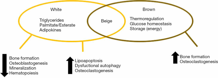
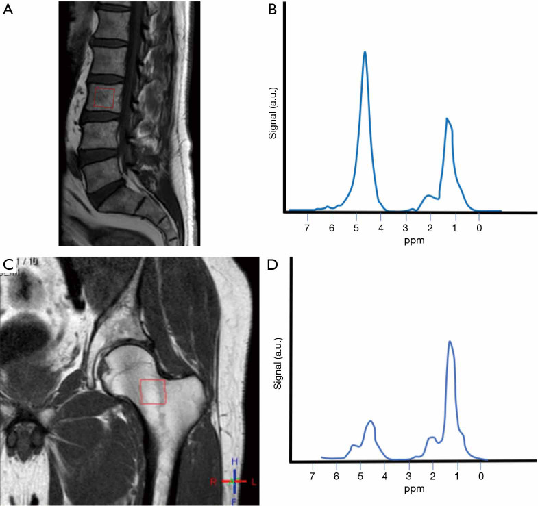
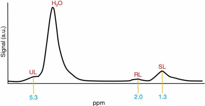
The study of bone has for many years been focused on the study of its mineralized component, and one of the main objects of study as radiology developed as a medical specialty. The assessment has until recently been almost limited to its role as principal component of the scaffolding of the human body. Bone is a very active tissue, in continuous cross-talk with other organs and systems, with functions that are endocrine and paracrine and that have an important involvement in metabolism, ageing and health in general. Bone is also the continent for the bone marrow, in the form of "yellow marrow" (mainly adipocytes) or "red marrow" (hematopoietic cells and adipocytes). Recently, numerous studies have focused on these adipocytes contained in the bone marrow, often referred to as marrow adipose tissue (MAT). Bone marrow adipocytes do not only work as storage tissue, but are also endocrine and paracrine cells, with the potential to contribute to local bone homeostasis and systemic metabolism. Many metabolic disorders (osteoporosis, obesity, diabetes) have a complex and still not well-established relationship with MAT. The development of imaging methods, in particular the development of cross-sectional imaging has helped us to understand how much more laid beyond our classical way to look at bone. The impact on the mineralized component of bone in some cases (e.g., osteoporosis) is well-established, and has been extensively analyzed and quantified through different radiological methods. The application of advanced magnetic resonance techniques has unlocked the possibility to access the detailed study, characterization and quantification of the bone marrow components in a non-invasive way. In this review, we will address what is the evidence on the physiological role of MAT in normal skeletal health (interaction with the other bone components), during the process of normal aging and in the context of some metabolic disorders, highlighting the role that imaging methods play in helping with quantification and diagnosis.

Keywords: Bone marrow; adipose tissue; body composition; magnetic resonance spectroscopy (MRS); osteoporosis.


Conflict of interest statement

Conflicts of Interest: All authors have completed the ICMJE uniform disclosure form (available at http://dx.doi.org/10.21037/qims.2020.01.11). The special issue “Body Composition Imaging” was commissioned by the editorial office without any funding or sponsorship. GG served as the unpaid Guest Editor of the special issue and serves as an unpaid editorial board member of Quantitative Imaging in Medicine and Surgery. AB served as the unpaid Guest Editor of the special issue. The authors have no other conflicts of interest to declare.

Figures

Figure 1
Figure 1
Representation of the combined roles of bone marrow fat. White-like phenotype characteristics reduce bone formation and hematopoiesis, with an increase in adipogenesis and bone destruction. Brown-like phenotype stimulates bone formation and osteoblastogenesis.
Figure 2
Figure 2
Mechanisms of marrow behavior during loading of the subchondral bone. (A) Diagram representing the open model. The marrow fat (yellow arrows) and water (blue arrows) are assumed to flow freely through pores between compartments as the bone is compressed, and therefore only the walls (trabeculae) will bear the compression stress (red dotted arrows). (B) Diagram representing the closed model. In the closed model, there is no free transit of fat or water (yellow and blue arrows respectively), because the viscosity of fat does not allow it to pass through trabecular pores. The volume of fat marrow in the compartments remains constant and the compression causes bulging of the walls. This means the transformation of a loading force into tensile forces (green dotted arrows), that dissipate in a descending pressure gradient.
Figure 3
Figure 3
Difference in the spectral curve between spine and hip. Fifty-four-year-old male. (A) Voxel placed in the body of L3 (red box); (B) the spectral curve shows abundant water (strong water peak) in comparison to lipids. Conversion into fatty marrow happens from appendicular to axial skeleton, and within the long bones, from diaphysis to metaphysis. In adults, yellow bone marrow is mainly located in the appendicular skeleton. When there is a strong water peak, the extraction of the spectrum of unsaturated lipids will be challenging; (C) same patient, voxel placed in the femoral neck (red box); (D) lipids peak is stronger in this location.
Figure 4
Figure 4
Proton magnetic resonance spectroscopy. Stimulated echo acquisition mode (STEAM) single-voxel 1H-MRS sequence. The spectrum that results shows peaks for water (H2O), saturated lipids (SL), unsaturated lipids (UL) and residual lipids (RL). Fat content is related to water content and expressed as a percentage, called bone marrow fat fraction (BMFF). This percentage is calculated using the large lipid peak at 1.3 parts per million (ppm), which are the SL, normally disregarding the smaller lipid peaks at 2.0 and 5.3 ppm, which correspond to RL and UL, but these can also be used. 1H-MRS, proton magnetic resonance spectroscopy.
Figure 5
Figure 5
Comparative spectra between a 43-year-old woman [A, T1 sagittal planning image with vertebral body voxel (red box); B, spectral curve] and a 42-year-old man [C, T1 sagittal planning image with vertebral body voxel (red box); D, spectral curve]. A very subtle difference in the height (signal) of the lipid peak (at 1.3 ppm) can be appreciated, when comparing woman (12.27) and man (13.43), with a slightly higher peak present in the spectrum for the man, and less total water (89.42 in woman, 48.49 in man). Fat fraction {calculated as fat content (%) = [I fat/(I fat + I water)] ×100, where I is signal amplitude} in the woman was calculated at 12.06%, and 21.6% in the man. Men have approximately 6–10% more vertebral MAT than women in the 20 to 60 age group. SL, saturated lipids; MAT, marrow adipose tissue.
Figure 6
Figure 6
Comparison of spectral readings in two subjects, with normal BMD and in the osteoporosis range. (A) T1 sagittal planning image with vertebral body voxel (red box) in a 65-year-old woman; (B) BMD obtained with DXA is in the normal range (T=−0.3); (C) representation of the obtained spectral curve, with an increase in the lipid peak, and a decrease in the water peak, when compared to young subjects; (D) T1 sagittal planning image with vertebral body voxel (red box) in another 65-year-old woman affected by osteoporosis; (E) BMD obtained by DXA is in the osteoporotic range (T=−2.6); (F) spectral reading shows a high peak of lipids, with substantial decrease of water contents. BMD, bone mineral density; DXA, dual-energy X-ray absorptiometry.

References

    1. Guerri S, Mercatelli D, Aparisi Gómez MP, Napoli A, Battista G, Guglielmi G, Bazzocchi A. Quantitative imaging techniques for the assessment of osteoporosis and sarcopenia. Quant Imaging Med Surg 2018;8:60-85. 10.21037/qims.2018.01.05 - DOI - PMC - PubMed
    1. Curtis E, Litwic A, Cooper C, Dennison E. Determinants of Muscle and Bone Aging. J Cell Physiol 2015;230:2618-25. 10.1002/jcp.25001 - DOI - PMC - PubMed
    1. Tagliaferri C, Wittrant Y, Davicco MJ, Walrand S, Coxam V. Muscle and bone, two interconnected tissues. Ageing Res Rev 2015;21:55-70. 10.1016/j.arr.2015.03.002 - DOI - PubMed
    1. Bonewald L. Use it or lose it to age: A review of bone and muscle communication. Bone 2019;120:212-8. 10.1016/j.bone.2018.11.002 - DOI - PMC - PubMed
    1. Ponti F, Guerri S, Sassi C, Battista G, Guglielmi G, Bazzocchi A. Imaging of diabetic bone. Endocrine 2017;58:426-41. 10.1007/s12020-017-1278-5 - DOI - PubMed