. 2020 Jul 29:36:22.
doi: 10.1186/s42826-020-00055-z. eCollection 2020.

Study of ethion and lipopolysaccharide interaction on lung in a mouse model

Affiliations

Study of ethion and lipopolysaccharide interaction on lung in a mouse model

Geetika Verma et al. Lab Anim Res. .

Abstract

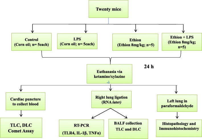
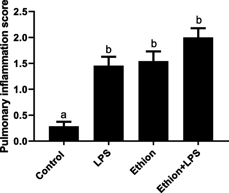
Ethion is an organophosphate used commonly in India despite being banned in many other countries. The present study was designed to study the interaction of ethion and lipopolysaccharide (LPS) together on lung after single low dose ethion exposure. Mice (n = 20) were alienated into control and treatment groups (n = 10 each). The treatment group was orally fed ethion (8 mg/kg/animal/day) dissolved in corn oil. The animals (n = 5 each) from both the groups were challenged with 80 μg Escherichia coli lipopolysaccharide (LPS) intranasally and the remaining animals (n = 5 each) were administered normal saline solution after 24 h. Ethion along with LPS induced lung inflammation as indicated by increased neutrophils and total leukocyte count (TLC) in broncheoalveolar lavage fluid. Ethion induced histomorphological alterations in lung as shown by increased pulmonary inflammation score in histopathology. Real time PCR analysis showed that ethion followed by LPS resulted significant (p < 0.05) increase in pulmonary Toll-like receptor (TLR)-4 (48.53 fold), interleukin (IL)-1β (7.05 fold) and tumor necrosis factor (TNF)-α (5.74 fold) mRNA expression. LPS co-exposure suggested synergistic effect on TLR4 and TNF-α mRNA expression. Ethion alone or in combination with LPS resulted genotoxicity in blood cells as detected by comet assay. The data suggested single dietary ethion exposure alone or in conjunction with LPS causes lung inflammation and genotoxicity in blood cells.

Keywords: Ethion; Genotoxicity; IL-1β; Organophosphates; TLR-4; TNF- α.


Conflict of interest statement

Competing interestsThe authors declared no potential conflicts of interest with respect to the research, authorship, and/or publication of this article.

Figures

Fig. 1
Fig. 1
Schematic diagram depicting the layout of the experiment
Fig. 2
Fig. 2
a TLC (× 103/μl) of blood, b nNeutrophil, c lLymphocyte (%) in blood; d TLC (× 103/ml) and e nNeutrophil (%) of BAL fluid after single dietary ethion exposure alone or in combination with LPS. a,b,c,d: no common superscript (a, b, c, d) between two levels of an effect indicates significant difference (p < 0.05) among groups
Fig. 3
Fig. 3
Haematoxylin and Eosin staining of Paraffin sections of lung showing normal histoarchitecture (A, B); peribronchial mononuclear infiltrating cells (arrow) and infiltration of inflammatory cells around alveolar septa (star) following treatment with LPS (C, D), ethion alone (E, F), or in combination with LPS (G, H). B: Bronchiole; PVS: Perivascular space; BV: Blood vessel; Original magnification A, C, E, G: 10 X; B, D, F, H: 40 X
Fig. 4
Fig. 4
Histopathological changes in lung expressed as pulmonary inflammation score after single dietary exposure to ethion for 24 h alone and with LPS. The results of five samples from each group are expressed as mean ± SE. Different superscript letters between two levels of an effect indicate significant difference (p < 0.05)
Fig. 5
Fig. 5
a Fold change expression of TLR4 b IL-1β and c TNFα mRNA after single ethion exposure for 24 h alone or along with LPS challenge. a,b,c:No common superscript between two levels of an effect indicates significant difference (p < 0.05)
Fig. 6
Fig. 6
Lung section showing immunopositive reaction in alveolar cells (single arrow), septal cells (double arrow) and endothelial cells of large blood vessel (arrow head). Representative images of TLR4, IL-1β, and TNF-α immunopositive cells after exposure to single ethion exposure and LPS. Original magnification: × 40
Fig. 7
Fig. 7
a Tail length (μm) and b tail DNA% after single ethion exposure with LPS. a,b,c,d: no common superscript between two levels of an effect indicates significant difference (p < 0.05) among groups

References

    1. Aktar MW, Sengupta D, Chowdhury A. Impact of pesticides use in agriculture: their benefits and hazards. Interdiscip Toxicol. 2009;2:1–12. doi: 10.2478/v10102-009-0001-7. - DOI - PMC - PubMed
    1. Verma G, Ramneek, Mukhopadhyay CS, Sethi RS. Acute ethion exposure alters expression of TLR9 in lungs of mice. Ind J Vet Anat 2016;28(1):40–43.
    1. Thakur J, Rao B, Rajwanshi A, et al. Epidemiological study of high cancer among rural agricultural community of Punjab in northern India. Int J Environ Res Public Health. 2008;5(5):399–407. doi: 10.3390/ijerph5050399. - DOI - PMC - PubMed
    1. Srivastava S, Narvi SS, Prasad SC. Levels of select organophosphates in human colostrum and mature milk samples in rural region of Faizabad district, Uttar Pradesh. India Human Exp Toxicol. 2011;30:1458–1463. doi: 10.1177/0960327110396525. - DOI - PubMed
    1. Dewan A, Patel AB, Pal RR, et al. Mass ethion poisoning with high mortality. Clin Toxicol (Phila) 2008;46:85–88. doi: 10.1080/15563650701517251. - DOI - PubMed

LinkOut - more resources