Review
. 2018 Oct 17:4:1-9.
doi: 10.1016/j.tcsw.2018.10.001. eCollection 2018 Dec.

Fission yeast cell wall biosynthesis and cell integrity signalling

Affiliations
Review

Fission yeast cell wall biosynthesis and cell integrity signalling

Pilar Pérez et al. Cell Surf. .

Abstract

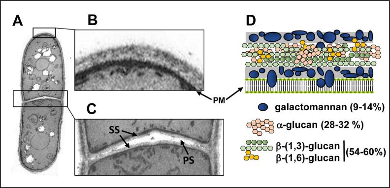
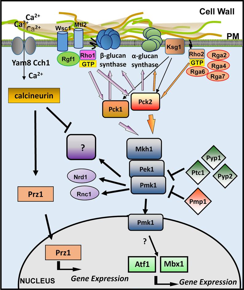
The cell wall is a structure external to the plasma membrane that is essential for the survival of the fungi. This polysaccharidic structure confers resistance to the cell internal turgor pressure and protection against mechanical injury. The fungal wall is also responsible for the shape of these organisms due to different structural polysaccharides, such as β-(1,3)-glucan, which form fibers and confer rigidity to the cell wall. These polysaccharides are not present in animal cells and therefore they constitute excellent targets for antifungal chemotherapies. Cell wall damage leads to the activation of MAPK signaling pathways, which respond to the damage by activating the repair of the wall and the maintenance of the cell integrity. Fission yeast Schizosaccharomyces pombe is a model organism for the study morphogenesis, cell wall, and how different inputs might regulate this structure. We present here a short overview of the fission yeast wall composition and provide information about the main biosynthetic activities that assemble this cell wall. Additionally, we comment the recent advances in the knowledge of the cell wall functions and discuss the role of the cell integrity MAPK signaling pathway in the regulation of fission yeast wall.

Keywords: Bgs; Cell wall; GTPase; MAPK; Pkc; Polysaccharides; β-glucan, α-glucan.


Figures

Fig. 1
Fig. 1
(A) TEM image of a fission yeast cell. Details of (B) the cell wall and (C) the septum are shown. PM: Plasma membrane, PS: primary septum, SS: Secondary septum. (D) Scheme of the fission yeast cell wall organization and percentages of the different polysaccharides.
Fig. 2
Fig. 2
Schematic representation of the β-(1,3)-glucan synthesis by the GS complex in the plasma membrane. Then the β-(1,3)-glucan is elongated and branched through the action of cell wall proteins with glucosyltransferase activity. Glucose units (formula image).
Fig. 3
Fig. 3
Scheme of Rho GTPases activation cycle. Top, GDP-bound Rho GTPases are activated by GEFs, which are proteins that promote the substitution of GDP by GTP. Active GTP-bound Rho GTPases promote the activation of different effectors. The intrinsic GTPase activity is stimulated by GAPs and causes the hydrolysis of the GTP into GDP and the return of the Rho GTPase to the GDP-bound inactive state. The GDI proteins extract GDP-bound Rho GTPases from the membrane. Bottom, the different S. pombe Rho GEFs and GAPs described to date are shown.
Fig. 4
Fig. 4
(A) Kite and Doolittle hydropathy plot and model of the Bgs proteins structure with 15–16 transmembrane domains. (B) Fluorescence micrographs showing the localization of GFP-Bgs1 in exponentially growing S. pombe cells. (C) Localization of GFP-Bgs4 in calcofluor-stained growing cells. (D) Kite and Doolittle hydropathy plot and model of the Ags1 protein structure. (E) Time-lapse micrographs showing Ags1-GFP in exponentially growing cells stained with calcofluor (lower panels). CW: calcofluor white. The bar is 5 μm.
Fig. 5
Fig. 5
Schematic representation of S. pombe cell wall integrity pathway. Rho1 is activated by the sensors Wsc1 and Mtl2, which detect and transmit signals coming from the cell wall through the GEF Rgf1. Rho1 activates y the β-(1,3)-glucan synthase and stabilizes the kinases Pck1 and Pck2. Both kinases are activated by the Phospholipid-dependent kinase Ksg1 and in turn activate the β-(1,3)-glucan synthase. Rho2 is not activated by Wsc1 or Mlt2, but regulates the α-(1,3)-glucan synthase via Pck2. Additionally, Rho2-Pck2 are the main activators of the MAPK cell integrity pathway. Signaling of this pathway is transmitted through the MAPK module to Atf1 and Mbx1 transcription factors in the nucleus and to different targets such as the RNA-binding proteins Rnc1 or Ndr1 in the cytoplasm. Pmk1 also activates the Yam8/Cch1 calcium channel that in turns activates the phosphatase complex calcineurin. This phosphatase antagonizes the function of MAPK pathway in response to several stimuli. Other phosphatases including Pmp1, and the SAP-dependent phosphatases Ptc1, Pyp1 and Pyp2 negatively regulate Pmk1 activation.

References

    1. Aimanianda V., Clavaud C., Simenel C., Fontaine T., Delepierre M., Latge J.P. Cell wall b-(1,6)-glucan of Saccharomyces cerevisiae: structural characterization and in situ synthesis. J. Biol. Chem. 2009;284:13401–13412. - PMC - PubMed
    1. Alfa C., Fantes P., Hyams J., McLeod M., Warbrick E., editors. Experiments with Fission Yeast: A Laboratory Course Manual. Cold Spring Harbor Laboratory Press; Cold Spring Harbor, N.Y.: 1993.
    1. Arasada, R., Pollard, T.D., 2014. Contractile Ring Stability in S. pombe Depends on F-BAR Protein Cdc15p and Bgs1p Transport from the Golgi Complex. Cell reports. - PMC - PubMed
    1. Arellano M., Duran A., Perez P. Rho 1 GTPase activates the (1–3)beta-D-glucan synthase and is involved in Schizosaccharomyces pombe morphogenesis. EMBO J. 1996;15:4584–4591. - PMC - PubMed
    1. Arellano M., Duran A., Perez P. Localization of the Schizosaccharomyces pombe rho1 GTPase and its involvement in the organization of the actin cytoskeleton. J. Cell Sci. 1997;110:2547–2555. - PubMed

LinkOut - more resources