Review
. 2019 Mar 8:5:100021.
doi: 10.1016/j.tcsw.2019.100021. eCollection 2019 Dec.

Nanomotion detection based on atomic force microscopy cantilevers

Affiliations
Review

Nanomotion detection based on atomic force microscopy cantilevers

A C Kohler et al. Cell Surf. .

Abstract

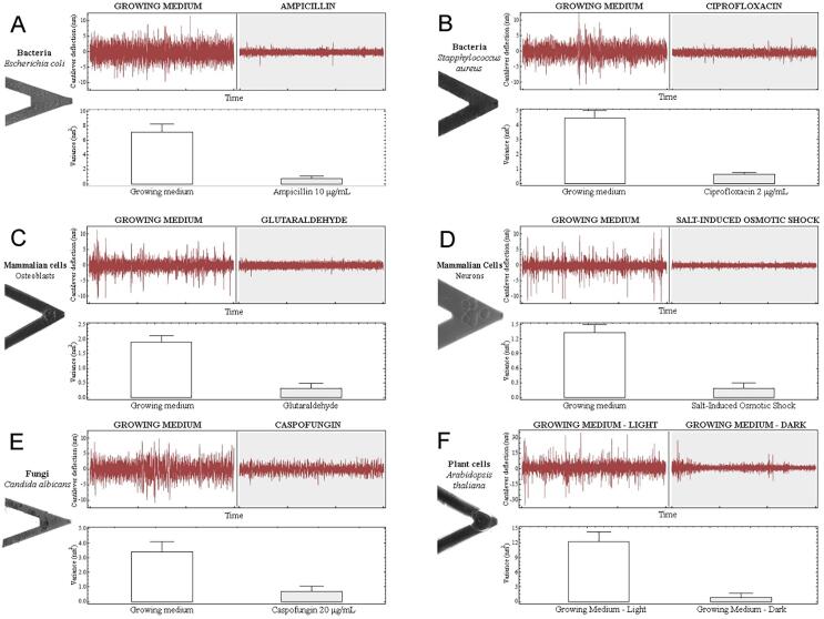
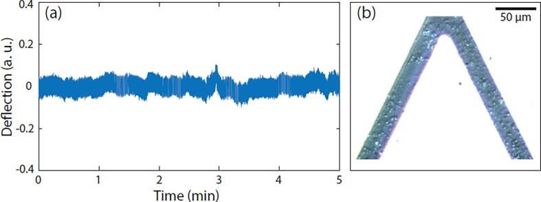
Atomic force microscopes (AFM) or low-noise in-house dedicated devices can highlight nanomotion oscillations. The method consists of attaching the organism of interest onto a silicon-based sensor and following its nano-scale motion as a function of time. The nanometric scale oscillations exerted by biological specimens last as long the organism is viable and reflect the status of the microorganism metabolism upon exposure to different chemical or physical stimuli. During the last couple of years, the nanomotion pattern of several types of bacteria, yeasts and mammalian cells has been determined. This article reviews this technique in details, presents results obtained with dozens of different microorganisms and discusses the potential applications of nanomotion in fundamental research, medical microbiology and space exploration.

Keywords: AFM; Antibiotic Susceptibility Test (AST); Cell viability; Nanomotion detection.


Figures

Fig. 1
Fig. 1
. Principle of AFM and nanomotion detection A: An AFM cantilever (3) is scanning a surface (4) with living organisms and their topographic characteristics are recorded by the means of the laser displacement (1) by a 4-quadrant photodiode (2). B: basic AFM-based nanomotion detection. The living organisms are attached onto the functionalized cantilever and its oscillations are followed as in a normal AFM procedure. C: Example of an in-house made nanomotion detector. 1: laser, 2: analysis chamber with AFM cantilever (not visible in this image), 3: optical microscope and CCD camera, 4: photodetector and preamplifier.
Fig. 2
Fig. 2
. Versatility of nanomotion to determine the viability of cells Nanomotion of several organisms were determined in different conditions. These graphs represent nanomotion of bacteria (A, B), fungi (E), mammalian (C, D) and plant cells (F) in growth media and after addition of molecules that alter their viability. In all the type of cells tested, the cantilever deflection as well as the variance of the cantilever deflection drastically decrease in presence of these specific molecules: antibiotics for bacteria (A, B), cross-linking agent or osmotic shock for C and D respectively, antifungal for E and exposure to darkness for plant cells. The picture of the cantilever highlights the presence of the cells throughout the experiment, providing evidence that the decrease is not due to loss of cells from the cantilever. Figure adapted from PNAS, January13,2015,112,2,378–391 with authorization.
Fig. 3
Fig. 3
. Mechanism susceptible to cause cell nanomotion A: several molecular and cellular mechanism could be involved in the nanomotion of a cell. Among them, mitochondria and other metabolic processes could generate motion of the overall cell. Cell membrane motion itself could be part of the numerous mechanisms causing cellular nanomotion. B: extra-cellular organelles – such as pili or flagella – are motile components used for the locomotion of cells or bacteria this inherently to its function causes nanomotion of cells or bacteria. C: ion channels (green) change conformation in order to let in or out specific ions (depicted as H + in yellow). This structure rearrangement affects the lipid bilayer of cells and could be part of the nanomotion of cells. (For interpretation of the references to colour in this figure legend, the reader is referred to the web version of this article.)
Fig. 4
Fig. 4
. Nanomotion signal induced by active mitochondria. Oscillations of the AFM cantilever as a function of time (a) with active mitochondria attached onto its surface (b). Reprinted from Stupar et al. (2017) Mech. Sci., 8, 23–28, 2017.

References

    1. Alonso-Sarduy L., De Los Rios P., Benedetti F., Vobornik D., Dietler G., Kasas S., Longo G. Real-time monitoring of protein conformational changes using a nano-mechanical sensor. PLoS One. 2014;9 e103674. - PMC - PubMed
    1. Binnig G., Quate C.F., Gerber C. Atomic force microscope. Phys. Rev. Lett. 1986;56:930–933. - PubMed
    1. Boisen, A., 2011. Cantilever-like micromechanical sensors.
    1. Braun T., Ghatkesar M.K., Backmann N., Grange W., Boulanger P., Letellier L., Lang H.-P., Bietsch A., Gerber C., Hegner M. Quantitative time-resolved measurement of membrane protein–ligand interactions using microcantilever array sensors. Nat. Nanotechnol. 2009;4:179–185. - PubMed
    1. Domínguez C.M., Kosaka P.M., Sotillo A., Mingorance J., Tamayo J., Calleja M. Label-free DNA-based detection of mycobacterium tuberculosis and rifampicin resistance through hydration induced stress in microcantilevers. Anal. Chem. 2015;87:1494–1498. - PubMed