Long-term follow-up of penile glans necrosis due to paraphimosis
- PMID: 32743402
- PMCID: PMC7292180
- DOI: 10.1002/iju5.12064
Long-term follow-up of penile glans necrosis due to paraphimosis
Abstract
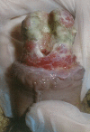
Introduction: Paraphimosis is a urologic emergency in which the foreskin of the penis becomes trapped behind the coronal sulcus and forms a tight band of constricting tissue. Surgical or conservative release of this constriction is required for the treatment. Delayed treatment will cause devastating outcomes, such as penile glans necrosis. A few studies have reported penile glans necrosis/gangrene, but long-term follow-up of the recovery from glans necrosis due to paraphimosis has not been previously reported.
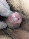
Case presentation: A 25-year-old man who experienced glans necrosis following paraphimosis was not treated promptly with circumcision. The patient underwent conservative treatment with debridement of necrotic tissue and cystostomy for urethral meatal necrosis. We were able to prevent partial penectomy. His penile glans was covered with healthy epithelium and retained its natural shape and voiding and erectile functions were normal 2 years after the treatment.
Conclusion: We report successful conservative management of penile glans necrosis.
Keywords: cystostomy; debridement; long‐term follow‐up; paraphimosis; penile necrosis.
© 2019 The Authors. IJU Case Reports published by John Wiley & Sons Australia, Ltd on behalf of the Japanese Urological Association.
Conflict of interest statement
The authors declare no conflict of interest.
Figures


References
-
- Hollowood AD, Sibley GN. Non‐painful paraphimosis causing partial amputation. Br. J. Urol. 1997; 80: 958. - PubMed
Publication types
LinkOut - more resources
Full Text Sources
